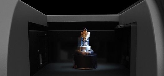
SKYSCAN 1272 CMOS Edition
A sharp 3D view of your sample


Highlights
SKYSCAN 1272 CMOS – High-resolution XRM
SKYSCAN 1272 CMOS Edition builds on the trusted SKYSCAN 1272 desktop X-ray Microscopy platform and incorporates the latest X-ray technologies to bring XRM to the next level.
The state-of-art 16 megapixels CMOS X-ray detector provides high-contrast images with superior resolution. The extended detector field of view and enhanced sensitivity for X-rays result in up to twice shorter scan times.
The SKYSCAN 1272 CMOS offers automatic selection of parameters with Genius-Mode. Magnification, energy, filter, exposure time and background corrections can all be optimized automatically with a single click.
A 16-position sample changer is optionally available for unattended high throughput scanning.
SKYSCAN 1272 CMOS is complemented by 3D.SUITE. This comprehensive software suite covers GPU-accelerated reconstruction, 2D/ 3D morphological analysis, as well as surface and volume rendering visualization.
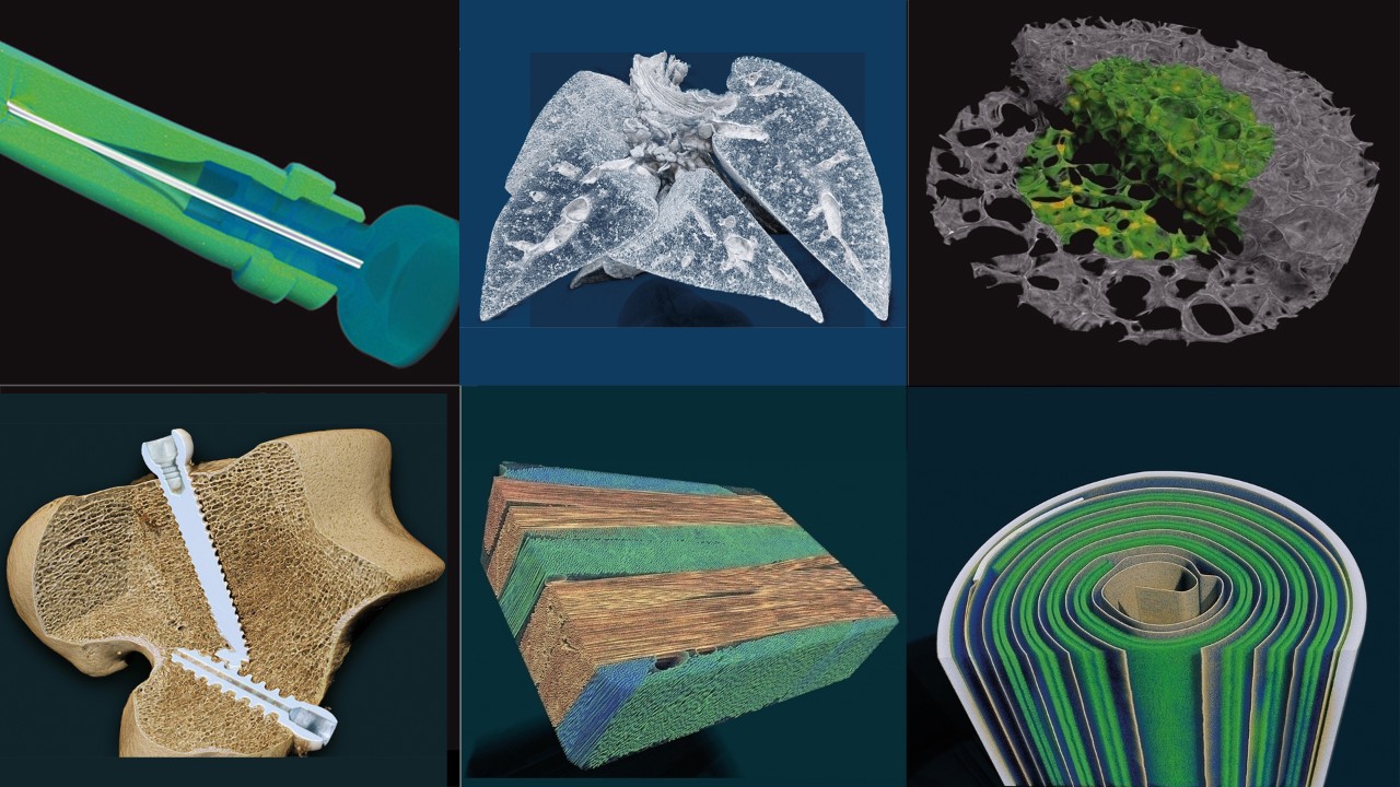
Market Gallery
Pharmaceuticals
Porous media
Building Materials
Electronic Components/Devices
Geological/ Petrochemical
Fibers/Composites
Key Features
Genius Mode
The SKYSCAN 1272 CMOS offers automatic selection of parameters with Genius-Mode. Magnification, energy, filter, exposure time and background corrections can all be optimized automatically with a single click.
Furthermore, both the sample and large-format CMOS camera can be positioned as close as possible to the source, which substantially increases the measured intensity. That is why SKYSCAN 1272 CMOS scans up to 5 times faster compared to conventional systems with fixed camera position.
Sample changer
SKYSCAN 1272 CMOS can optionally be equipped with an external 16-position sample changer to increase throughput for QC and routine analysis.
The sample changer accepts a variety of sample sizes, up to a diameter of 25 mm.
Samples can be can be easily replaced at any time without interrupting an ongoing scanning process. New samples are automatically detected, and LED‘s indicate the status for every scan: ready, scanning, done.
In-situ stages
The Bruker material testing stages are designed to perform compression experiments up to 4400 N and tensile experiments up to 440 N. All stages automatically communicate through the system’s rotation stage, without the need of any cable connections. Using the supplied software, scheduled scanning experiments can be set up.
Bruker's heating and cooling stages can reach temperatures of up to +80ºC, or 30ºC below ambient temperature. Just like the other stages, no extra connections are needed, and there is an automatic recognition of the stage. Using the heating & cooling stages, samples can be examined under non-ambient conditions, to evaluate the effect of temperature on the sample’s microstructure.
SKYSCAN 1272 CMOS
No external cooling water or special power with no compromise in performance: Designed for the ecological and economical needs of today
SKYSCAN 1272 CMOS
Smart solutions and design, such as the integrated vibration isolation, form a perfect whole
Highlighted Applications
Fibers and Composites
By combining materials into a composite the resulting component can have increased strength while significantly decreasing weight. Further optimization comes from ensuring the orientation of the subcomponents are optimized. One of the classic components used are fibers ranging from steel rebar in concrete, glass fibers in electronic components, to carbon nanotubes in aviation materials. XRM allows inspection of fibers and composites without the need for cross sectioning, ensuring the condition of the sample is not affected by sample preparation.
- Orientation of embedded objects
- Quantification of layer thickness, fiber sizes and separation
- In-situ temperature and physical properties testing with accessory stages
Pharmaceuticals
Development of a new pharmaceutical is a time consuming and costly endeavor. XRM can accelerate time-to-market by providing immediate feedback on the product’s internal structure during the product formulation stage.
- Determine the table compaction density
- Measure coating thickness uniformity
- Evaluate API distribution
- Detect stress induced micro-cracks in compacted tables
- Apply in-situ compression for testing the mechanical properties
Foams
Foams are widely used for industrial applications. Whether they are used as thermal or acoustic insulators, shock absorbers in protective parts, filters … depends on the material and the structural properties of the foam.
XRM enables to visualize a foam’s 3D internal structure in a non-destructive way.
- Determine the structural thickness of local struts
- Determine the structure separation to visualize the pore network
- Apply in-situ mechanical tests with compression and tensile stages
- Quantify the level of open and closed porosity
Application Gallery
Electronics
Geology
Wood
Food
Porous Structures
SKYSCAN 1272 CMOS Specifications
Feature |
Specification |
Benefit |
X-ray source |
40 – 100 kV 10 W < 5 µm spot size at 4 W |
Maintenance-free sealed X-ray source Fast scans for QC, or 4D XRM |
X-ray detector |
16 MP sCMOS detector (4096 x 4096 pixels) | Fine-pitched detectors for achieving highest resolution |
Object size |
75 mm diameter 80 mm height |
Capable to scan from small to medium large sample sizes |
Sample changer (optional) |
16 samples up to 25 mm diameter External access |
Unattended high throughput Any combination of large and small samples Add/remove samples at any time without interrupting the actual scan |
Dimensions |
W 1160 mm x D 520 mm x H 330 mm Weight 150 kg |
Space-saving desktop system that fits in every lab |
Power supply |
100-240V AC, 50-60Hz, 3A max. |
Minimum installation requirements, a standard power supply suffices |
POSITION, SCAN, RECONSTRUCT and ANALYZE
Bruker XRM solutions include all software needed to collect and analyze data. An intuitive graphical user interface with user guided parameter optimization support both expert and novice users. By using the latest GPU powered algorithms, reconstruction time is substantially reduced. CTVOX, CTAN and CTVOL combine to form a powerful suite of software for both qualitative and quantitative analysis of models.
Measurement Software:
SKYSCAN 1272 – Instrument control, measurement planning and collection
Reconstruction Software:
NRECON – Transforms the 2D projection images into 3D volumes
Analysis Software:
DATAVIEWER – Slice-by-slice inspection of 3D volumes and 2D/3D image registration
CTVOX – Realistic visualization by volume rendering
CTAN – 2D/3D image analysis & processing
CTVOL – Visualization of surface models to export for CAD or 3D printing
SKYSCAN 1272 Resources
Brochures & Flyers
Videos
Service & Support
- Helpdesk for technical issues with hardware, software, and applications support using web based and advanced remote service tools.
- LabScape Maintenance Service Agreements
- On-site, on-demand support
- Installation and operational qualification as well as performance verification
- Site planning, relocation, and consultation
- Replacement and spare parts, consumables, and in-person and online training
- Software updates, manuals, and LabScape MSA management (↗brukersupport.com)

