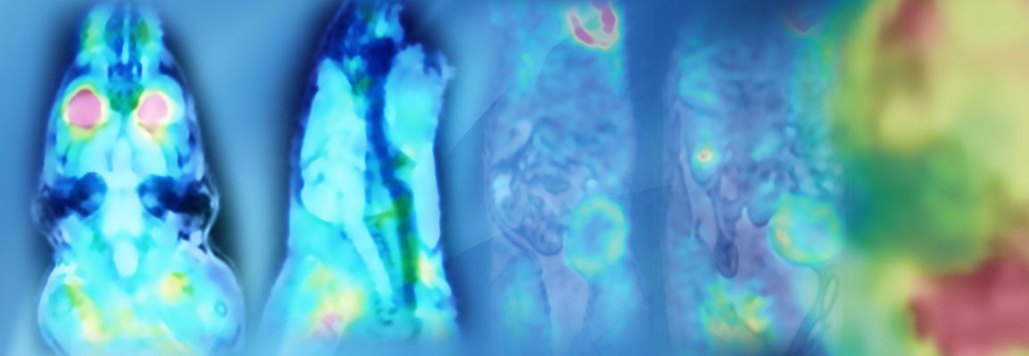

Early Detection of Metabolic Remodeling in Pressure Overload Left Ventricle Hypertrophy using Dynamic PET Imaging
In this webinar, Professor Bijoy Kundu will describe the application of PET imaging for understanding the early metabolic changes that arise in animal models of hypertension-induced LVH and heart failure. Kundu and team have developed dynamic/kinetic imaging techniques and obtaining time-resolved images from PET scanners to assess the metabolic changes that occur in the heart when it is subjected to pressure overload. Clinicians might be interested in using PET to detect metabolic changes in patients with early stages of heart failure, with a view to prevent the structural changes that are seen later with MRI.
This webinar took place on December 13th, 2018
What to Expect
Dr. Kundu will review the imaging modalities applied in his lab, with a focus on Bruker’s Albira trimodal PET∕SPECT∕CT scanner for cardiovascular imaging. The projects the group has been working on will be described and a basic understanding of what happens to the heart when it is under stress will be given and how novel PET imaging methods could contribute to new early diagnosis and treatments.
Key Points
- The use of PET for longitudinal imaging of early metabolic changes in LVH
- The advantages of using PET to assess disease progression
- The hypothesis that metabolic changes in the heart may precede and even trigger structural and functional remodeling
- The potential application of PET to identify these changes early on to prevent remodeling and heart failure in the long term
Who Should Attend?
Basic scientists, research assistants and other academics who already use PET, as well as those working with MRI but who are curious about PET, would all benefit from attending. Clinicians in academia and diagnostics who are interested in imaging disease, particularly the dynamic imaging of cardiovascular disease.
Speaker
Dr. Bijoy K. Kundu
Associate Professor - Department of Radiology and Medical Imaging University of Virginia School of Medicine
Bijoy K. Kundu, PhD, is an Associate Professor in the tenure track in the department of Radiology and Medical Imaging at the University of Virginia, Charlottesville. After completing his PhD in Nuclear Physics from the Bhabha Atomic Research Center, India, he pursued his post-doctoral work at the Indian Institute of Technology, Kanpur, India. He then moved to the Department of Radiology, University of Virginia, as a post-doctoral associate and moved up the faculty ranks. Dr. Kundu is a member of many scientific societies and reviewer of a number of peer-reviewed journals. The goals of his lab are to develop and optimize quantitative cardiac PET imaging techniques to address the hypothesis of metabolic remodeling in small animal models of myocardial injury and type 2 diabetes. His lab is currently funded by a grant from the National Institutes of Health. https://med.virginia.edu/radiology-research/bijoy-kundu/