Light-Sheet Fluorescence Microscopy for Living Plants
Advancing Plant Imaging with Light-Sheet Fluorescence Microscopy
Light-sheet fluorescent microscopy (LSFM) is well-suited to overcome many of the complications around imaging live and delicate plants. Because of this capability, researchers use LSFM to create compelling data about plants through innovative image acquisition and data processing.
This application note, “Light-Sheet Fluorescence Microscopy for Living Plants,” discusses the importance of using light-sheet fluorescence microscopy to overcome many of the challenges associated with imaging plants. Learn about the technical components of a light-sheet system – including light-sheet thickness and aperture – and data processing solutions for creating compelling data. Real-world examples and figures also explain how to use these components to study plant dynamics (growth) and structures (cell walls and tissues).
Contents include:
KEYWORDS: Plant Biology; Case Study; Light-Sheet Fluorescence Microscopy; Image Acquisition and Data Analysis
Introduction
The study of plants is not only important to support our understanding of biological mechanisms, but also to foster knowledge of crop improvement and safety, especially with respect to the global economy.1 While plant samples were traditionally studied as thin dyed sections, 3D in situ live imaging is the current state-of-the-art research method. In recent years, the study of in situ 3D morphogenesis using fluorescence microscopy has been greatly enriched by the availability of a wide range of fluorescent tissue-specific reporter lines1 as well as genetically encoded biosensors.2
One major challenge in working with 3D plant tissue is that tissue is often inhomogeneous. This is further complicated by the fact that plant cells have a cell wall and chlorophyll containing tissues that absorb light, which limits the microscopy approaches that can be used.3 Additionally, plants have an intrinsic geometry with aerial parts that have natural tropisms for light and roots that are influenced by gravity. Thus, the sample setup does not only need to satisfy the physiological conditions (e.g., light or temperature) but also physical ones (e.g., large sample size and upright/vertical mounting).4
From an imaging point of view, fluorescence microscopy raises complications with the fact that lasers emit photons at several orders of magnitude higher than the sunlight to which plants are typically exposed.5 This can potentially impact photosynthesis, local temperature, and light exposure, all of which can lead to sample deterioration or imaging artifacts.1,4
An elegant solution to the above problems is light-sheet fluorescence microscopy (LSFM). LSFM uses a sheet of light for selective plane excitation while information is collected simultaneously by an uncoupled emission objective and a camera.6 This setup means that large samples can be studied as the beam paths can penetrate the tissue. However, only a thin sheet is illuminated so phototoxicity is low, and most LSFM setups allow for vertical sample embedding and full environmental control, which is critical for sample health.
In this application note we highlight challenges with using microscopy for image acquisition of plants, challenges specific to LSFM, data processing challenges, and how Bruker Luxendo can support you in overcoming these obstacles to create meaningful data.
Challenges of Plant Imaging
Capturing Dynamic Processes
Most biological processes are inherently dynamic so the ability to acquire data over time is often better than single timepoint acquisition with only a snapshot of information. However, live imaging is a trade-off between preserving the sample and acquiring good data with sufficient signal quality and resolution.4
Visualizing Tissues
Despite the increasing availability of tissue-specific fluorescent markers, dyes are still often used to visualize tissues of interest. Unfortunately, dyes can bleach upon laser exposure and some can become potentially toxic.2 That is why reducing photons with techniques such as LSFM is pivotal for sample health.
Aerial Parts vs Roots
Studying plants beyond the sprouting phase means having to consider aerial parts and roots. Aerial parts contain chlorophyll, which not only reacts to light exposure, but scatters light. Furthermore, chloroplasts, the cells containing chlorophyll, are autofluorescent and give off even additional fluorescence.1 Roots, especially root tips, are often studied in morphogenesis, but they strongly react to gravitational forces and growth geometry can change rapidly. Thus, using adaptive microscopy approaches is needed for timelapses longer than 30 minutes.2
Light-sheet fluorescence microscopes are made with a variety of components that make them well-suited to overcome these considerations. Voluminous sample chambers provide space for the samples to grow, vertical embedding considers gravitation, and environmental control creates consistent conditions for samples. To improve data quality, images can be acquired from multiple views (e.g., Bruker’s MuVi SPIM) that are subsequently merged to derive data with high signal and resolution. To address sample movement and/or growth outside the desired field of view (FOV), adaptive autofocus can help with automatic sample tracking.
Cell Walls
Like an exoskeleton, cell walls provide mechanical and chemical resistance for plants and contain 40%-to-60% cellulose, 10%-to-24% lignin, and varying amounts of other cell wall proteins. This composition makes them a complex 3D network where cellulose fibrils can further trigger light scattering.7
However, as cell walls have a 3D geometry, the ability to image living plants in situ greatly assists with sample preparation challenges such as plant section quality, or finding the correct position and FOV of interest in a section.7
Challenges of LSFM
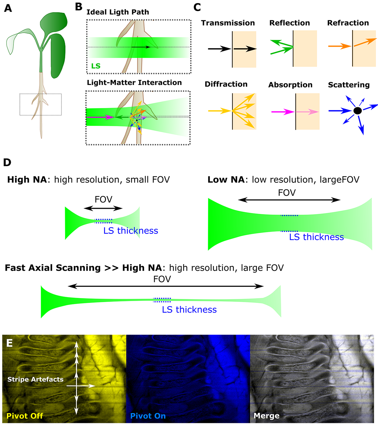
Light Interaction with Matter
Light interacts with matter, which leads to light ray interference by changes in transmission, reflection, refraction, diffraction, absorption, and scattering.8,9 The impact of matter on light rays can be particularly pronounced when the refractive indices of sample, embedding medium, and materials are mismatched. One way to optimize the optical properties of the system is the use of a low numerical aperture (NA) illumination objective and a high NA detection objective. This allows for a thin light-sheet with wide-field detection.5
Since artifacts, such as shadowing and striping, are particularly pronounced in static singleside illumination setups, a pivoted the light-sheet and multi-view approach can help reduce these detrimental issues. Lastly, lattice LSFM combined with structured illumination techniques can further improve data resolution.10
Light-Sheet Thickness
In LSFM, the light sheet is central to everything. The thickness of the sheet determines not only the amount of sample that is illuminated, but also the thinner the sheet, the higher the resolution in z. In combination with fast axial scanning, the entire FOV is homogenously illuminated. Additionally, the beam geometry is important, especially with respect to placing the beam waist in the centre of the FOV.11
The MuVi SPIM produces a homogenously illuminated FOV using fast axial scanning. This works by sweeping a tightly focused Gaussian beam along the illumination axis, leading to an elongated, uniform, and thin light sheet that provides uniform axial resolution over large FOVs.
Multi-View Imaging
Data can be acquired from multiple views and angles, and subsequently fused to increase data quality and reduce artifacts. Importantly, the fusion can be done with or without fiduciary markers. Together with the ability to cover large specimens with high-quality images, this can also provide isotropic spatial resolution.12
The MuVi SPIM allows for sample rotation, enabling a 360° view and making it the fastest multi-angle view system on the market. The next-generation MuVi SPIM also shows an unrivalled signal-to-noise ratio due to light-sheet scanning, adjustable light-sheet thickness, robust aberration tolerance, and dual de-striping via pivot scanning.
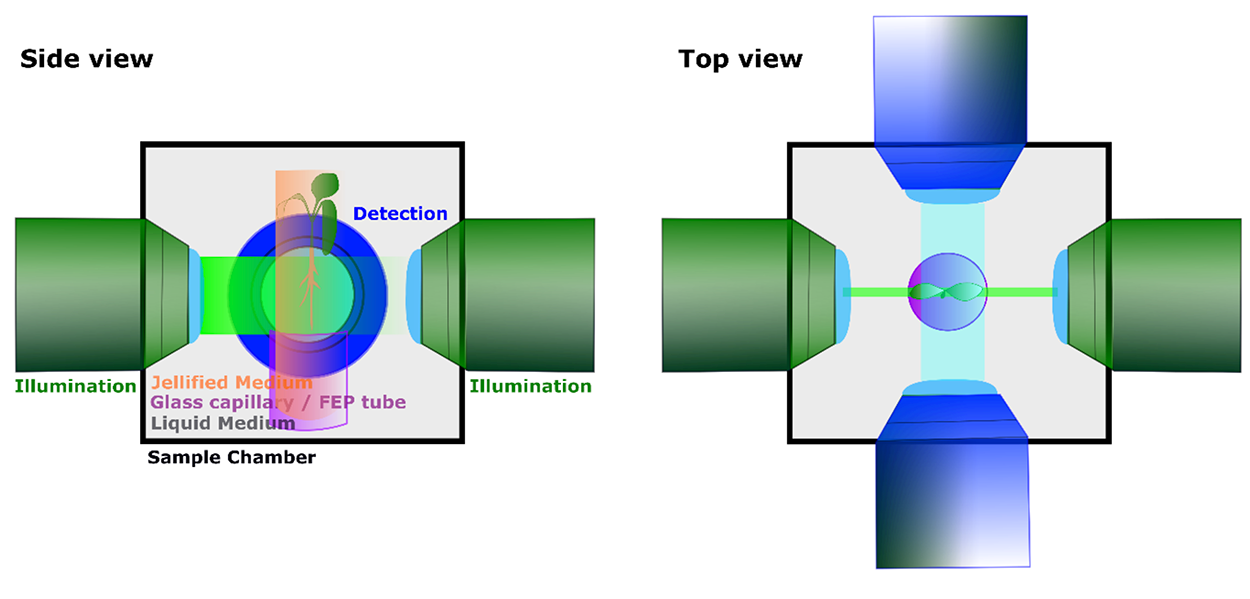
Flexibility in Setup and Embedding
Most LSFM systems allow for vertical sample embedding, as well as sample chamber access. Depending on what stage and organ of the plant one wants to study, several embedding options exist. For example, one can use jellifying media (e.g., phytogel, low melting agarose) and holders (e.g., glass capillary or FEP tubes) to establish a top-holding or bottom-holding setup.3 Environmental control can be established in the sample chamber and held stable over time. Additionally, the sample chamber can be filled with medium, which can be exchanged over time to avoid wearing-off or building-up of compounds making use
Lastly, it is often useful to apply more advanced imaging techniques or transiently perturb the system to observe biological behavior. This allows novel insights into the biology of processes.4 To achieve this, Bruker Luxendo has light-sheet solutions for photomanipulation (PM module) and advanced imaging (AIM – advanced imaging module) techniques.
Data Challenge
Handling Data Analysis with Luxendo Software
In this era of data, one major challenge is the analysis and storage of acquired data, particularly LSFM, where datasets can easily be in the terabyte range.13 Bruker’s LuxBundle allows for effective image registration and fusion, which enables data to be processed with application-specific software solutions.
To ensure long-term data storage and archiving, Luxendo Bruker also offers the well-known ACQUIFER HIVE solution. To effectively analyze data, application specific programs are being developed to aid image plant cell segmentation and analysis, such as lineage tracing, (e.g., MorphographX,14 PlantSeg,15 or MARS/ALT16).
Application Example
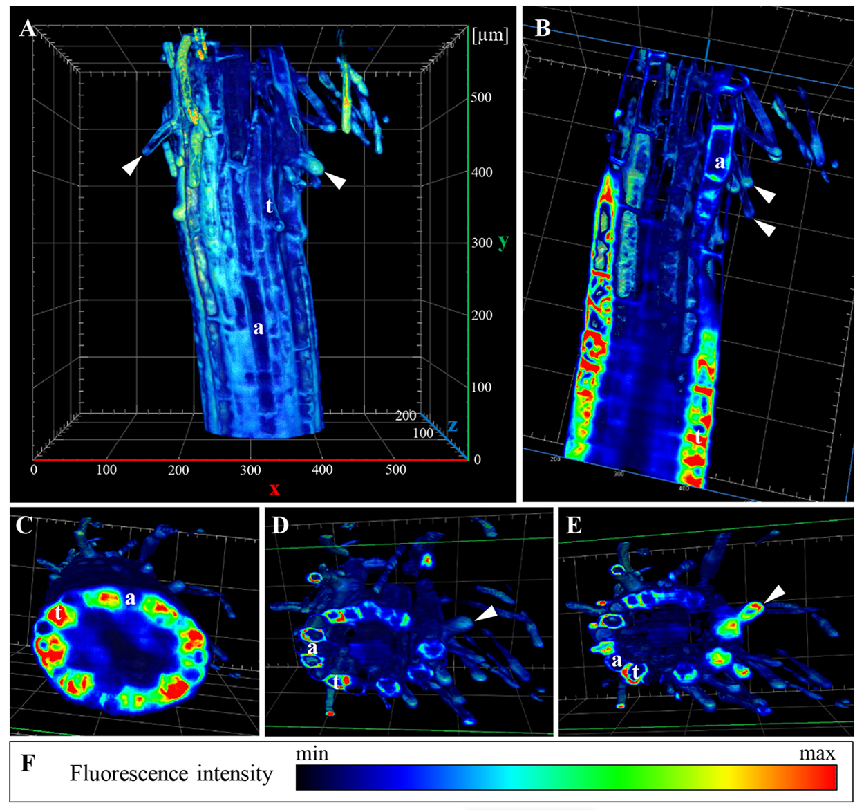
LSFM to Study ANNEXIN 1 in Arabidopsis
Annexins are an evolutionary conserved superfamily of multifunctional proteins which are fundamental to many processes, such as cell growth, differentiation, and stress responses. In this case study, Ticha et al. produce a transgenic Arabidosis line to visualize endogenous ANNEXIN 1 (ANN1) and study its in vivo distribution using LSFM.17
Conclusion
Imaging living plants involves challenges on several levels, including sample properties (e.g., aerial and roots, chlorophyll, cell walls), tissue visualization (e.g., dyes vs reporter lines), sample-friendly imaging (e.g., embedding, low phototoxicity, timelapses), and efficient data handling (e.g., processing and storage). Working together with end users and application specialists, Bruker Luxendo has the experience needed to deliver expert commercial LSFM solutions that can significantly aid the imaging of living and naturally growing plants.
Authors
Elisabeth Kugler, MarCom Specialist (E.Kugler-extern@bruker.com);
Monika Löschinger, Senior Product and Application Scientist
(Monika.loschinger@bruker.com);
Malte Wachsmuth, Director (Malte.wachsmuth@bruker.com);
Melissa Martin, Life Science Writer (Melissa.martin@bruker.com)
References
- M. Ovecka et al., “Imaging plant cells and organs with light-sheet and super-resolution microscopy,” Plant Physiology, vol. 188, no. 2, pp. 683–702, Feb. 2022, doi: 10.1093/plphys/kiab349.
- L. Colin et al., “Imaging the living plant cell: From probes to quantification,” The Plant Cell, vol. 34, no. 1, pp. 247–272, Jan. 2022, doi: 10.1093/plcell/koab237.
- B. Berthet and A. Maizel, “Light sheet microscopy and live imaging of plants,” Journal of Microscopy, vol. 263, no. 2, pp. 158–164, 2016, doi: 10.1111/jmi.12393.
- G. V. Reddy, S. P. Gordon, and E. M. Meyerowitz, “Unravelling developmental dynamics: transient intervention and live imaging in plants,” Nat Rev Mol Cell Biol, vol. 8, no. 6, Art. no. 6, Jun. 2007, doi: 10.1038/nrm2188.
- E. H. K. Stelzer, “Light-sheet fluorescence microscopy for quantitative biology,” Nat. Methods, vol. 12, no. 1, pp. 23–26, Jan. 2015, doi: 10.1038/nmeth.3219.
- J. Huisken, J. Swoger, F. Del Bene, J. Wittbrodt, and E. H. K. Stelzer, “Optical sectioning deep inside live embryos by selective plane illumination microscopy,” Science, vol. 305, no. 5686, pp. 1007–1009, Aug. 2004, doi: 10.1126/science.1100035.
- G. Costa and I. Plazanet, “Plant Cell Wall, a Challenge for Its Characterisation,” Advances in Biological Chemistry, vol. 6, no. 3, Art. no. 3, May 2016, doi: 10.4236/abc.2016.63008.
- J. Ryan, A. R. Gerhold, V. Boudreau, L. Smith, and P. S. Maddox, “Introduction to Modern Methods in Light Microscopy,” in Light Microscopy: Methods and Protocols, Y. Markaki and H. Harz, Eds. New York, NY: Springer, 2017, pp. 1–15. doi: 10.1007/978-1-4939-6810-7_1.
- E. Kugler and E. Reynaud, “LSFM series – Surfing on the data freak wave! Part II: Before imaging: Know your sample (geometry),” FocalPlane, Oct. 10, 2020. https://focalplane.biologists.com/2020/10/10/lsfm-series-surfing-on-the-data-freakwave-part-ii-before-imaging-know-your-sample-geometry/ (accessed Nov. 16, 2022).
- P. J. Keller et al., “Fast, high-contrast imaging of animal development with scanned light sheet-based structured-illumination microscopy,” Nat Methods, vol. 7, no. 8, pp. 637–642, Aug. 2010, doi: 10.1038/nmeth.1476.
- M. Weber, M. Mickoleit, and J. Huisken, “Light sheet microscopy,” Methods Cell Biol., vol.123, pp. 193–215, 2014, doi: 10.1016/B978-0-12-420138-5.00011-2.
- R. K. Chhetri, F. Amat, Y. Wan, B. Höckendorf, W. C. Lemon, and P. J. Keller, “Wholeanimal functional and developmental imaging with isotropic spatial resolution,”
- A. Schlaeppi et al., “Meeting in the Middle: Towards Successful Multidisciplinary Bioimage Analysis Collaboration,” Frontiers in Bioinformatics, vol. 2, 2022, Accessed: Apr. 19, 2022. [Online]. Available:https://www.frontiersin.org/article/10.3389/fbinf.2022.889755
- P. Barbier de Reuille et al., “MorphoGraphX: A platform for quantifying morphogenesis in 4D,” eLife, vol. 4, p. e05864, May 2015, doi: 10.7554/eLife.05864.
- A. Wolny et al., “Accurate and versatile 3D segmentation of plant tissues at cellular resolution,” eLife, vol. 9, p. e57613, Jul. 2020, doi: 10.7554/eLife.57613.
- R. Fernandez et al., “Imaging plant growth in 4D: robust tissue reconstruction and lineaging at cell resolution,” Nat Methods, vol. 7, no. 7, Art. no. 7, Jul. 2010, doi: 10.1038/nmeth.1472.
- M. Tichá et al., “Advanced Microscopy Reveals Complex Developmental and Subcellular Localization Patterns of ANNEXIN 1 in Arabidopsis,” Frontiers in Plant Science, vol. 11, 2020, Accessed: Jan. 10, 2023. [Online]. Available: https://www.frontiersin.org/articles/10.3389/fpls.2020.01153
©2023 Bruker Corporation. ACQUIFER HIVE, LuxBundle, Luxendo, and MuVi are trademarks of Bruker. All other trademarks are the property of their respective companies. All rights reserved. AN2010, Rev. A0.