
Optical approaches for investigating neuromodulatory effects on amygdala dynamics and behavior
Unraveling neuronal ensembles with deep brain calcium imaging
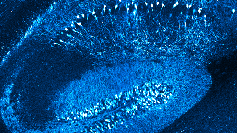
In this webinar, Dr. Piantadosi showcases his use of multiphoton optogenetics to manipulate optically separable amygdalar valence encoding ensembles in vivo. He also discusses how to overcome the challenges associated with using this technique deep in the brain.
Presenter’s Abstract
A long-standing theory postulates that monoaminergic neuromodulatory inputs control the gain of neuronal ensembles and that this motif is well suited to govern valence processing that occurs in limbic brain regions, including the basolateral amygdala (BLA). However, direct experimental evidence for how neuromodulatory input alters neural ensemble gain and the cell types and receptors that participate is not clear. Using deep brain calcium imaging approaches and focusing on the highly conserved noradrenergic input from the locus coeruleus (LC) to the amygdala, I have tried to understand how neuromodulatory input contributes to changes in correlated activity of amygdala ensembles.
I find that activation of this circuit promotes anxiety-like behavior, and this change in behavior coincides with an increase in synchronous activity of large groups of BLA neurons in a β-adrenergic receptors (β-AR) dependent manner. This increased connectivity may reflect a change in network gain. To test the functional consequence of increased ensemble synchrony, I have pioneered the use of multiphoton optogenetics in the deep brain. Using 2-photon calcium imaging and multiphoton stimulation I have found that synchronous activation of amygdala ensembles defined by their valence specificvalence-specific activity profiles can bias behavioral output, demonstrating a causal role for this gain control mechanism. Further, we use this approach to investigate the connectivity of these opposing ensembles in vivo, identifying functional antagonism between ensembles.
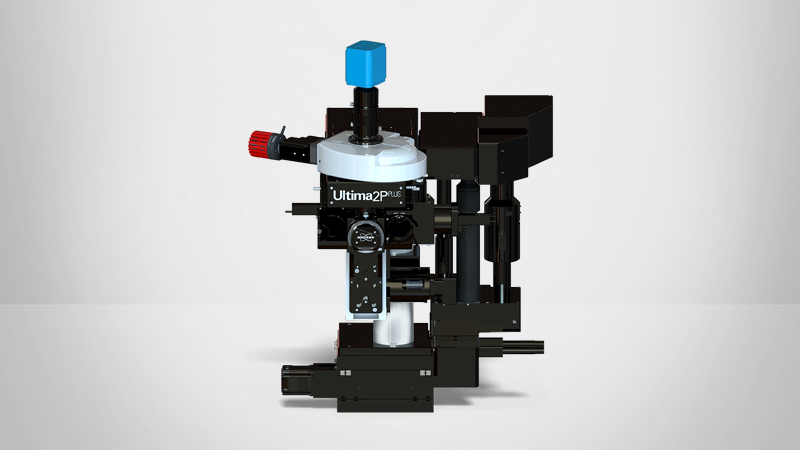
Find out more about the technology featured in this webinar or our other solutions for Multiphoton Microscopy:
Featured Products and Technology
Speaker
Sean Piantadosi, Ph.D.,
Postdoctoral Scholar, University of Washington


