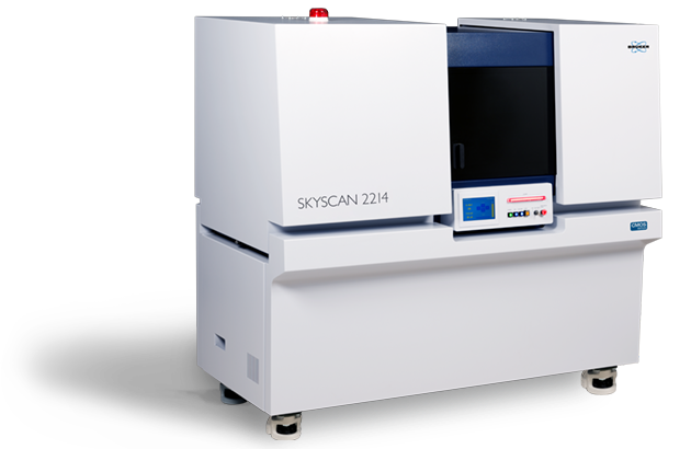
SKYSCAN 2214 CMOS Edition
Bigger. Brighter. Bolder.


Field of View, Resolution, Power
Watch the videos to get more insight
SKYSCAN 2214 CMOS – Nanoscale X-ray Microscopy
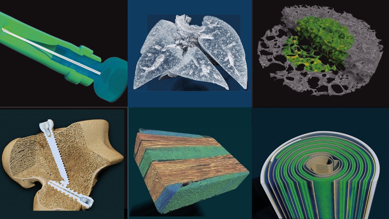
The MULTISCALE X-ray microscope SKYSCAN 2214 CMOS covers the widest range of object sizes and spatial resolutions in a single instrument, enabling advanced 3D imaging and exact modeling of geological materials in oil and gas exploration, composite materials, Li batteries, fuel cells, electronic assemblies, as well as ex-vivo preclinical applications like in lung imaging or tumor vascularization.
The instrument allows scanning and 3D non-destructive reconstruction of the internal microstructure of objects as large as >300mm in diameter as well as submicron resolution for small samples.
The system is equipped with an "open type" transmission X-ray source with <0.5 micron spot size and a diamond window. It can accommodate up to four X-ray detectors for maximum flexibility. Automatic variable acquisition geometry and phase-contrast enhancement allows best possible quality in relatively short scanning time.
SKYSCAN 2214 is complemented by 3D.SUITE. This comprehensive software suite covers GPU-accelerated reconstruction, 2D/ 3D morphological analysis, as well as surface and volume rendering visualization.
Market Gallery
Key Features
Source
The SKYSCAN 2214 uses a latest generation open-type X-ray source. The source offers true spatial resolution below 500 nm, an X-ray energy up to 160 keV and source power up to 16 W. The source is practically maintenance-free with an extremely easy pre-aligned filament replacement procedure.
The SKYSCAN 2214 has an open-type (pumped) nanofocus
X-ray source with diamond window. It produces an X-ray beam with peak
energy from 20 kV to 160 keV and is supplied with two types of cathodes.
The tungsten (W) cathodes operate in the full range of accelerating
voltages up to 160 kV and provide a spot size down to 800 nm. The
lanthanum hexaboride (LaB6) cathodes can be used for accelerating
voltages from 20 kV to 100 kV and provide a spot size of the X-ray beam
smaller than 500 nm to achieve the highest resolution in imaging and 3D
reconstruction. The JIMA resolution pattern indicates that 500 nm
structures can be easily resolved.
For long-term stability of the focal spot size and position of the emission point, the X-ray source is equipped with a liquid cooling system which contains a re-circulator providing precise temperature stability of the cooling fluid.
Detectors
The SKYSCAN 2214 can be equipped with up to four X-ray cameras for ultimate flexibility: three CMOS cameras with different resolution and field of view balance, and one flat panel detector to cover an XL field of view. All cameras can be selected with just a single mouse click.
Using large-format CMOS detectors with small pixel size allows extension of high-resolution 3D imaging to large objects. The built-in detector flexibility enables adjusting the field of view and spatial resolution according to the object size and density. An advanced reconstruction from a volume of interest provides scanning of a selected part of a large object with high resolution without compromising image quality.
Additionally, the field of view can be increased horizontally and vertically by using offset camera positions and vertical object movement. The 3D.SUITE software automatically stitches the different images together with compensation of the shifts and possible intensity differences
As research topics and analytical needs evolve, cameras can be retro-fitted at any point of time during the system’s lifetime.
In-situ stages
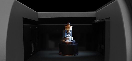
The high-precision object stage of the SKYSCAN 2214 supports objects up to 300 mm diameter and 20 kg in weight. The air-bearing rotation motor allows precise rotation of objects at very high accuracy, and the integrated micro-positioning stage guarantees a perfect sample alignment.
The SKYSCAN 2214 has a large and easily accessible sample chamber to allow scanning of big objects as well as mounting of optional stages. On top plenty of space is available for peripheral equipment.
The Bruker material testing stages are designed to perform compression experiments up to 4400 N and tensile experiments up to 440 N. All stages automatically communicate through the system’s rotation stage, without the need of any cable connections. Using the supplied software, scheduled scanning experiments can be set up.
Bruker's heating & cooling stages can reach temperatures of up to +80 °C or 30 °C below ambient temperature. Just like the other stages, no extra connections are needed, and there is an automatic recognition of the stage. Using the heating & cooling stages, samples can be examined under non-ambient conditions, to evaluate the effect of temperature on the sample’s microstructure.
The SKYSCAN 2214 is fully compatible with stages from DEBEN. With the included adapter, the DEBEN stage can be simply placed onto the rotation stage of the SKYSCAN 2214.
Large Chamber
Push-button Detector Change
Advanced Source
Micro-Positioning Sample Stage
Helical Scanning
HART Plus
Highlighted Applications
Additive manufacturing
Additive manufacturing, also commonly called 3D printing, allows the creation of components with complex external and internal structure. Unlike classic techniques which require special molding or tooling, additive manufacturing allows the economical production of both one piece prototypes and large batch production parts. Once completed, confirmation of both the internal and exterior structure is important in ensuring that the component will perform as intended. XRM allows this inspection in a non-destructive manner, giving confidence that a component will meet or exceed specifications.
- Inspection of internal voids for trapped powder
- Validation of external and internal dimensions
- Direct comparison with CAD models
- Analysis of single and multi-material components
Fibers and composites
By combining materials into a composite the resulting component can have increased strength while significantly decreasing weight. Further optimization comes from ensuring the orientation of the subcomponents is optimized. One of the classic components used are fibers ranging from steel rebar in concrete to carbon nanotubes in aviation materials. XRM allows inspection of fibers and composites without the need for cross-sectioning, ensuring the condition of the sample is not affected by sample preparation.
- Orientation of embedded objects
- Quantification of layer thickness and fiber sizes
- In-situ temperature and physical properties testing with accessory stages
Geology
The study of geological specimens, whether it is a core sample from deep below the surface or a rock laying on the ground, offers a wealth of information into the formation of the world around us. Analysis often requires destruction of the pristine sample, removing important provenance of the internal structures. XRM gives a view into the sample without sectioning, allowing faster time to result and the possibility of future analysis.
- Density dependent 3D visualization of the specimen interior
- Pore network visualization
- Digital sectioning allowing application standard geological methods
SKYSCAN 2214 Specifications
Feature | Specification | Benefit |
X-ray source | 20-160 kV 16 W max. | User exchangeable filament Optimize for max power (W) or max resolution (LaB6) Rotatable diamond window for maximum lifetime |
X-ray detector | 6 Mp active pixel flat-panel 16 Mp large-format CMOS 16 Mp Mid-format CMOS 15 Mp High-res CMOS | Variety of pixel and detector sizes allow balance between detector resolution, coverage and counting statistics Available with 1, 2, 3 or 4 detectors Field upgradable to add detectors |
Image Formats | Up to 8000 x 8000 x 2300 pixels after a single scan | User selectable image size allows balance of dataset size and needed resolution Software allows downsizing after data collection |
Resolution | 60 nm smallest pixel size | Simple graphics control for optimizing experiment resolution based on selected detector, sample and detector distance Adjustable source focus size to balance maximum power and resolution |
Positioning Accuracy | <50 nm for rotation | Air bearing sample stage provides smooth rotation Simple chuck style mounting of sample posts Mechanical and electrical interface for advanced materials research stages |
Maximum Object Size | 300 mm in diameter (140 mm scanning size) 400 mm in length Maximum object weight 20 kg | Both the power and room to scan large samples Precise positioning of small samples near the source to maximize magnification |
| Dimensions | W 1800 mm x D 950 mm x H 1680 mm Weight: 1500 kg | Efficiently designed to optimize use of lab space Source maintenance access via interlocked large sliding door |
POSITION, SCAN, RECONSTRUCT and ANALYZE
Bruker XRM solutions include all software needed to collect and analyze data. An intuitive graphical user interface with user guided parameter optimization support both expert and novice users. By using the latest GPU powered algorithms, reconstruction time is substantially reduced. CTVOX, CTAN and CTVOL combine to forma powerful suite of software for both qualitative and quantitative analysis of models.
Measurement Software:
SKYSCAN 2214 – Instrument control, measurement planning and collection
Reconstruction Software:
NRECON – Transforms the 2D projection images into 3D volumes
Analysis Software:
DATAVIEWER – Slice-by-slice inspection of 3D volumes and 2D/3D image registration
CTVOX – Realistic visualization by volume rendering
CTAN – 2D/3D image analysis & processing
CTVOL – Visualization of surface models to export for CAD or 3D printing
Service & Support
- Helpdesk for technical issues with hardware, software, and applications support using web based and advanced remote service tools.
- LabScape Maintenance Service Agreements
- On-site, on-demand support
- Installation and operational qualification as well as performance verification
- Site planning, relocation, and consultation
- Replacement and spare parts, consumables, and in-person and online training
- Software updates, manuals, and LabScape MSA management (↗brukersupport.com)

