
BioAFM & NanoIR: New Insights into Nanomechanical and High-Resolution Chemical Analysis
Learn about leading-edge life science and biomedical research with AFM and AFM-IR
In this webinar, our expert guest speakers provide insights into their research using AFM and AFM-IR techniques, which includes the investigation of drug adsorption on metal carrier nanoparticles and characterization of virus-like particles.
Webinar Summary
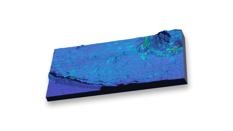
Atomic Force Microscopy (AFM) is an advanced multi-parametric imaging technique that enables the 3D imaging of the surface topography of living biological samples in the nm-range, the characterization of nanomechanical properties, and the visualization of structural changes occurring at the molecular level.
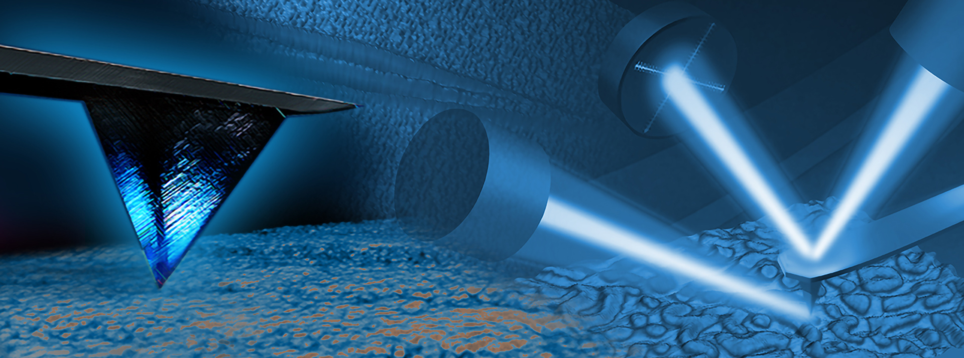
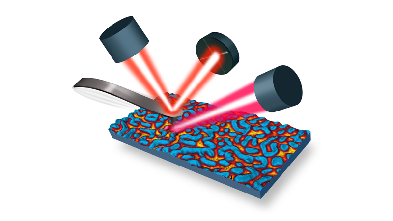
Nanoscale Infrared AFM spectroscopy and imaging (AFM-IR, which can be conducted on Bruker nanoIR platforms) measures spatially varying physical and chemical properties with nanoscale spatial resolution in samples as diverse as polymers, thin films, monolayers, and biological materials.
In this webinar, our expert guest speakers provide insights into their work using these techniques and speak on exciting applications in the field of life science and biomedical research. You can expect from this webinar to:
- Gain new insight into the capabilities and cutting-edge life science applications of AFM and AFM-IR techniques
- Explore the potential future uses and opportunities for discovery via both technologies
- Hear experts answer audience questions about the technical parameters, instrumentation, and results of their research
Find out more about the technology featured in this webinar or our other solutions for nano-IR or AFM for life science:
| Presentation Length | Presentation Title | Presenter | Abstract |
| 25 min | The Drug/Metal Nanocarrier Connection: Characterization and Imaging Using Nanoscale Infrared Spectroscopy | Dr. Natalia Piergies | Nowadays, nanotechnology plays an important role in improving conventional therapies, in particular in the treatment of cancer. Drug delivery systems, based on nanosized vehicles, deliver therapeutic agents to the target site where they are released in a controlled manner. The critical step in designing effective drug/carrier conjugates is the formation of a stable connection between the drug and the carrier surface. Therefore, monitoring how the drug deposits onto the carrier and understanding precisely how it is attached to the carrier`s surface is of great importance. Application of surface-enhanced vibrational spectroscopy methods enables the detailed interpretation of the drug/metal nanocarrier connection. In this webinar, Dr. Natalia Piergies discusses the surface enhancement occurring in the AFM-IR technique and its use in the nanoscale imaging of drug/metal nanocarrier conjugates. This is a groundbreaking approach, as even subtle changes in drug adsorption behavior can be tracked at very high spatial resolution. |
| 20 min | Probing Bio-Interfaces using Atomic Force Microscopy and Complementary Techniques | Dr. Aaron Elbourne | Atomic force microscopy allows in situ nanoscale systems to be probed unlike many other techniques. In this talk, Dr Aaron Elbourne speaks about his recent use of the Bruker JPK - BioAFM (NanoWizard 4) in tandem with other high-resolution imaging techniques. Specifically, the use of AFM techniques to probe fungal biofilms, virus-like-particles (VLPs), nanoparticle-cell interactions, and cell-virus adhesion are discussed. In addition, hyperspectral Synchrotron-based IR-spectroscopy are highlighted as a complimentary technique to these studies. |
Featured Products and Technology


Speakers

Natalia Piergies, Ph.D., Assistant Professor, Department of Experimental Physics of Complex Systems, Institute of Nuclear Physics PAN
Dr. Natalia Piergies is a specialist in surface-enhanced vibrational spectroscopy. She obtained her Ph.D. in 2014 from the Jagiellonian University. She joined the Department of Experimental Physics of Complex Systems at the Institute of Nuclear Physics PAN as an Assistant Professor in 2016.
Her scientific interest is focused on the application of metal nanoparticles as drug carriers which may improve the effectiveness of cancer therapies. She investigates the relationship between the drug/nanocarrier connection and the activity of these conjugates in in-vitro models.

Aaron Elbourne, Ph.D., Postdoctoral Research Fellow, School of Science, RMIT University
Dr. Aaron Elbourne is a postdoctoral research fellow within the School of Science at RMIT University. He currently holds a Jack Brockhoff Foundation Early Career Medical Research Fellowship and is a leader within RMIT’s ECR network. He obtained his Ph.D. in Chemistry in 2017 from The University of Newcastle, Australia under the supervision of Professor Erica J. Wanless. He began his postdoctoral fellowship in February of 2017. His early research focused on molecular-resolution atomic force microscopy (AFM) imaging, with an emphasis on fundamental ion adsorption at the solid-liquid interface.
His current research has 'shifted gears' focusing on anti-microbial surface and particle technologies and bio-interfacial studies. He has a passion for research with real-world applications and industrial translation. More broadly, he is interested in developing next-generation vaccine technologies, antimicrobial technologies, anti-cancer antibodies, and new methods for combating antibiotic resistance.