
Monitoring Radiotherapy Dose Distribution Using EPR Spectroscopy
“…EPR may play a future role for the investigation of dose distributions within sub-millimeter surface layers at tissue-air interfaces”
Radiotherapy is an important aspect of cancer treatment, shown by the fact that it is used in the management of around half of all cancer cases. It is considered to be the most effective strategy for reducing the risk of tumor regrowth after surgical excision.
Radiotherapy involves directing a beam of ionizing radiation at the tumor to kill the cancer cells. Since the radiation is also damaging to healthy cells, it is important that during radiotherapy high doses of ionizing radiation are specifically targeted at the tumor whilst minimizing exposure to the surrounding healthy cells in order to avoid side effects.
Targeted radiotherapy is generally achieved using imaging to ensure that the radiation beam is accurately directed at the cancer cells. Although many imaging modalities are available to provide such guidance, magnetic resonance imaging (MRI) has become the imaging tool of choice.
This offers benefits over other imaging tools in that it does not expose the patient to additional radiation and provides excellent soft-tissue contrast. Radiotherapy instrumentation is now available that performs MRI scanning in addition to delivering the therapeutic radiation.
However, there is evidence that the application of the magnetic field required for MRI scanning can interfere with the delivery of ionizing radiation to the target site. The radiation dose can be either increased or decreased as the trajectory of released electrons in the radiation beam is deflected by the magnetic field lines.
The exact extent of this effect has been difficult to quantify, but it appears to be especially significant at tissue-air interfaces due to the electron return effect (ERE).
Since such a disturbance has the potential to both reduce the efficacy of radiotherapy and increase the risk of side effects, considerable effort has been put into quantifying the extent of magnetic field induced perturbations in radiation dose distributions. However, quantifying the effect within a sub-millimeter thick surface layer has proved highly challenging.
Although simulation models have been used to theoretically assess the extent of radiation dose distribution perturbations, experimental validation of the findings continued to be sought. Some success was achieved using radiochromic film and plastic, but this is limited by the formation of artifacts that reduce the maximum surface-to-volume ratio of dosimeters.
The latest research using electron paramagnetic resonance (EPR) spectroscopy to quantify the magnetic field induced perturbations of radiation dose distributions indicates that this is indeed a feasible approach. EPR spectroscopy effectively measured magnetic-field induced perturbations of radiation dose distributions, even within a sub-millimeter thick surface layer at the dosimeter-air interface.
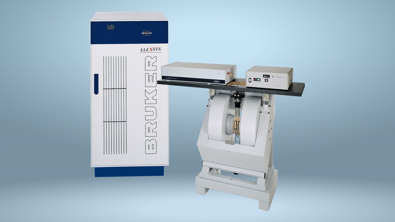
A Bruker ELEXSYS E580 X-band EPR spectrometer was used in pulsed mode to evaluate a 0.5 mm layer surrounding millimeter-sized air cavities for paramagnetic defects after irradiation with a 6 MV photon beam in both the presence and absence of a transverse magnetic field. The magnetic field gradient was generated in two planes using a Bruker E 540 GCX2 gradient coil system. The Bruker ER 4108 TMHS imaging resonator was used for echo detection.
The data revealed local dose enhancements and reductions of up to 35% and were in good quantitative agreement with the values derived theoretically using simulation models.
Reference:
Höfel S, et al. EPR-Imaging of magnetic field effects on radiation dose distributions around millimeter-sized air cavities. Physics in Medicine and Biology. 2019. DOI: 10.1088/1361-6560/ab325b/meta.


