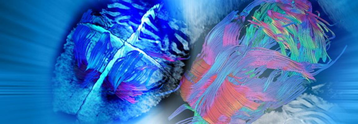

DTI connectomics in mouse models of neurological disorders
Since the typical length scale of water diffusion in tissue during an MRI experiment is around 1 µm, DTI can reveal structural information well below the nominal image resolution using biophysical modeling of the signal. In this Webinar, Dr. Philipp Boehm-Sturm will review the basics of diffusion imaging, as well as introduce the use of DTI connectomics for characterization of animal models of disease.
Dr. Boehm-Sturm will review protocol adaptation as well as good practice. He will then introduce sets of methods to reconstruct fiber tracts from DTI data and subsequent network analysis, the so-called DTI connectomics. Finally, from this information, structural and physiological parameters of the brain such as axone degradation, loss of tissue integrity or demyelinization can be elucidated. This particular approach will be demonstrated to characterize a mouse model of vascular cognitive impairment.
This webinar took place on March 28th, 2019
Key Topics
- Basics of diffusion imaging on Bruker scanners
- How to set up diffusion experiments
- Specificities of diffusion imaging, the dos and don´ts
- Process diffusion data to analyze the connectome of the mouse brain
- How to elucidate physiological information in the mouse brain from diffusion data using fiber tracking.
Who should attend?
Any scientist, research assistant or other academics with routine MRI knowledge who want to know more about diffusion imaging, as well as people who wish to get more out of their MRI system.
Speaker
Dr. Philipp Boehm-Sturm
Leader of the “Experimental MRI” group and scientific head of the Charité Core Facility „7 T experimental MRIs“ of the Charité University Medicine of Berlin, Germany
Philipp Boehm-Sturm, PhD, is heading a group and the core facility “7T experimental MRIs” at the Charité University Medicine Berlin, in Germany. After completing a PhD in the physics department of the University of Cologne and the Max-Planck-Institute for Neurological Research in Cologne, he transferred to the Experimental Neurology Department and the Center for Stroke Research at the Charité in Berlin to offer his expertise in small animal MRI and quickly became the scientific lead of the imaging core facility. Dr. Boehm-Sturm won several prestigious prices, including poster prices at the ESMRMB, EMIM, and the Congress of Stem Cells and Tissue Formation, and is involved in the organization of MRI conferences such as the Fluorine MRI Symposium 2017 in Berlin. Key areas of his expertise include neuroimaging, connectome of the mouse brain, fluorine MRI as well as alternative contrast agent development for MRI.