
microCT Applications in Lung Research and Immunology
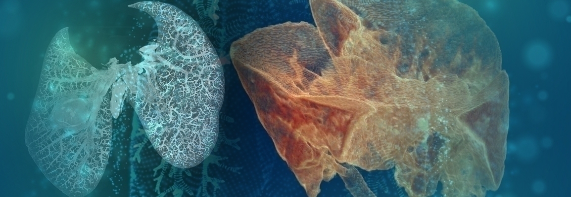
In this webinar, Dr. Elizabeth Redente, Associate Professor at National Jewish Health, will give insights from the literature and her group on the ability of microCT imaging to quantitate, visualize and follow the development of a myriad of lung diseases including inflammation associated with infection, fibrosis, emphysema and tumor development. The use of 3-dimensional analysis to quantify lung disease is advantageous over traditional histology techniques that capture a single cross-sectional analysis or macro-dissection of the lung to isolate tumors, which reduces spatial understanding. In addition, the use of contrast agents allows for easy visualization and quantitation of organ vasculature.
This webinar took place on May 28th, 2020
Who to Expect
In this webinar, Dr. Redente will discuss the advantages of microCT and provide a broad overview of the many pulmonary diseases that this imaging modality can visualize. This is a highly innovate technique and can be used to analysing living animals as well as fixed tissue. Examples from the literature will be provided as well as work from Dr. Redente’s laboratory.
Key Topics
microCT of lung
- Current methods and limitations
- Use of contrast agents
Examples of lung diseases
- Fibrosis
- Infections disease and inflammation
- Emphysema
- Lung cancer
Quantitation and visualization
- Lung volumes
- Hounsfield Units
- Emphysema
- Tumor counting
Benefits
- Higher differentiation of lung tissue and airspace
- High resolution images
- Low radiation dosage for longitudinal scans
- 3D reconstruction and visualization
Who Should Attend?
Researchers (undergraduates, graduates, postdoctoral fellows) in biomedical sciences who are interested in a gaining a better understanding of µCT techniques as well as an overview of current µCT research.
Speakers
Dr. Elizabeth Redente
Associate Professor National Jewish Health