
Light-Sheet Microscopy in the Study of Embryogenesis
Gain insights into mitotic chromosome organization with light-sheet microscopy
During this webinar, guest speaker Marina Makharova from EMBL Heidelberg, Germany discusses her lab’s use of Bruker’s light-sheet microscopy for real-time imaging of mitotic chromosomes in early mouse embryos. Hear about techniques for both visualizing and quantifying changes in the dimension and organization of mitotic chromosomes.
Presenter’s Abstract
The first embryonic divisions in mammals are surprisingly error-prone, leading to spontaneous abortion, congenital disease, and limited fertility. In non-mammalian model organisms, it has been shown that mitotic chromosomes adapt to the dramatic reduction in cell size with each cleavage division of the embryo - a process known as mitotic chromosome scaling, which has been hypothesized to protect genome integrity. However, it is unknown if chromosome scaling occurs in mammals and what the molecular mechanism is that shortens mitotic chromosomes in early embryos.
In this study, we imaged mitotic cells in preimplantation embryos to measure changes in mitotic chromosome dimensions during the first divisions after fertilization. In addition, we quantitatively analyzed the key protein complexes most likely to drive shortening, i.e., Condensin loop extruders, using single-molecule fluctuation calibrated confocal imaging. Our results show that mammalian embryos exhibit pronounced chromosome scaling and that early embryonic chromosomes are bound by much higher amounts of Condensin loop extruders compared to somatic cells.
Our quantitative imaging assays using light sheet microscopy will allow us to gain a mechanistic understanding of mitotic chromosome scaling in the future, by combining high-throughput, real-time imaging of live embryos with acute perturbation of the activity of Condensins.
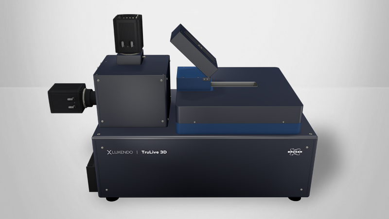
Find out more about the technology featured in this webinar or our other solutions for Light-Sheet Microscopy:
Featured Products and Technology
Guest Speakers
Marina Makharova, EMBL Heidelberg, Germany
The focus of Marina Makharova’s research is on understanding the principles of mitotic chromosome organization. After completing her Master's at Skoltech Institute, Moscow, she joined Jan Ellenberg’s Group (Cell division and nuclear organisation) at EMBL, Heidelberg, Germany, to work on mitotic chromosome compaction in pre-implantation mouse embryos. She combines various imaging techniques to characterize mitotic chromosome architecture and identify key players in mitotic chromosome scaling in cleavage-stage mammalian embryos.


