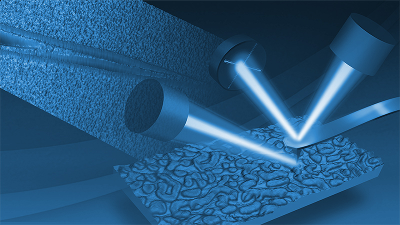
Resonance Enhanced Force Volume AFM-IR (REFV AFM-IR)
REFV AFM-IR extends the capabilities of nanoscale IR microscopy by combining force volume imaging with resonance-enhanced AFM-IR detection
Simultaneously collecting nanoscale chemical information and characterizing nanomechanical properties has always been of strong interest for atomic force microscopy. This application note introduces a method which realizes this goal by combining two established methods into one: Bruker’s Resonance Enhanced photothermal AFM-IR and force volume imaging. Implementation, operation, and application examples of this novel photothermal AFM-IR method are discussed in this application note, highlighting that the technique offers an accurate, accessible, and easy-to-use means for combined quantitative chemical and mechanical characterization.
Readers can expect to find:
- Background information framing the significance of Bruker’s new REFV AFM-IR technique
- Working principles descriptions for both Resonance Enhanced AFM-IR and force volume imaging, focusing on what benefits each imparts to REFV AFM-IR
- Detailed information on how REFV AFM-IR works, how it overcomes some common limitations, and what benefits it provides
- Four case studies, illustrating operation both with a fixed laser pulse rate and with a frequency sweep of laser pulse rate
KEYWORDS: Nanoscale Infrared Spectrometers; Nanoscale IR Spectroscopy; nanoIR; AFM-IR; AN207; Bruker; Application Note; Dimension IconIR
Introduction
Simultaneously collecting nanoscale chemical information and characterizing nanomechanical properties has always been of strong interest for atomic force microscopy. This application note introduces a method which realizes this goal by combining two established methods into one: Resonance Enhanced photothermal AFM-IR and force volume imaging. Implementation, operation, and application examples of this novel photothermal AFM-IR method are discussed in this application note, highlighting that the technique offers an accurate, accessible, and easy-to-use means for combined quantitative chemical and mechanical characterization
Background
Nanoscale infrared (nanoIR) microscopy enables label-free chemical imaging and spectroscopy at the nanometer scale by combining atomic force microscopy (AFM) with infrared radiation. Over the last years, AFM-IR has been developed with different AFM operational modes: Resonance Enhanced AFM-IR mode and the recently developed Surface Sensitive AFM-IR technique are based on contact mode, while Tapping AFM-IR is built on tapping mode.1 PeakForce Tapping®–based PeakForce infrared (PFIR) microscopy has lately joined as another AFM-IR mode.2 All these nanoIR variations inherit the advantages and limitations of their respective AFM base mode.
In this application note, we introduce Resonance Enhanced Force Volume (REFV) AFM-IR, a novel force volume–related approach with adjustable hold (dwell) time during which resonance-enhanced AFM-IR imaging or spectroscopy is performed. As shown in Figure 1, REFV AFM-IR offers the capability to perform simultaneous multimodal imaging collecting both mechanical properties such as elastic modulus and adhesion together with chemical information.
REFV AFM-IR provides:
- The capability to measure fragile, soft, sticky, or otherwise difficult samples with <10 nm spatial resolution and monolayer detection sensitivity by eliminating lateral forces
- Simultaneous nanomechanical and nanochemical measurements
- New, easy-to-use capabilities for artifact-free imaging and spectroscopy, decoupling mechanical property variations from AFM-IR data either with a frequency sweep during the dwell time or with a more traditional phase-locked loop (PLL)
- Simultaneous extraction of contact resonance data to deliver large datasets with complete sample information in one scan
Working Principles
Resonance Enhanced AFM-IR
IR signal detection in REFV AFM-IR utilizes the sensing concept of Resonance Enhanced AFM-IR. In Bruker’s patented Resonance Enhanced AFM-IR mode, the AFM cantilever is held on the sample in contact mode, and the laser pulse repetition rate is tuned to match a contact resonance frequency of the cantilever. Absorption of infrared radiation by the sample causes thermal expansion, which results in continuous excitation of the cantilever.
A Resonance-Enhanced AFM-IR spectrum is obtained by plotting the amplitude of a selected cantilever resonance in the frequency domain as a function of the laser wavelength. For both spectroscopy and imaging, a higher cantilever eigenmode (>1 MHz) provides higher spatial resolution and a smaller probing depth due to the shorter thermal diffusion length.
Using resonance-enhanced detection provides high sensitivity due to the enhancement of the AFM cantilever oscillation at resonant excitation. This allows for AFM-IR measurements on thin samples, such as self-assembled monolayers. The spatial resolution of Resonance-Enhanced AFM-IR can extend down to 20 nm for relatively smooth samples.3 To compensate for the mechanical difference between different components of the sample, a phase-locked loop (PLL) is usually implemented to maintain the match between the laser pulse rate and the local contact resonance frequency of the cantilever on the sample, thereby avoiding artifacts induced by mechanical variations of mechanical properties.3,4
Force volume imaging
A simple force curve taken with an AFM records the force felt by the tip as it approaches and retracts from a point on the sample surface. Force curves from an array of x-y points are combined into a three-dimensional array, or “volume,” of position-dependent force data (force volume), which can be used to extract quantitative nanomechanical properties such as adhesion, stiffness, or elastic modulus in real time for each sample location.
Force ramp rates up to 300 Hz (FASTForce Volume™, FFV) are possible using Bruker’s proprietary low-force trigger capability with a relative force setpoint measured against the probe deflection when not yet contacting the sample and thus removing deflection drifts inherent in the absolute trigger of contact mode. A dwell or hold segment, inserted into each approach-retract cycle, can serve to extract complementary spectral information. For example, advanced quantitative nanomechanical methods such as the FFV CR mode (contact resonance mapping) and AFM-nDMA (nanoscale dynamic mechanical analysis) use a short dwell segment during which viscoelastic properties are computed from a contact resonance spectrum or force modulation spectrum, respectively.5,6 The DataCube hyperspectral electrical modes form another example: an electrical spectrum (for example I-V curves in DataCube TUNA or C-V ones in DataCube SCM) is extracted during the dwell time, by ramping the sample voltage.7
How REFV AFM-IR works
In REFV AFM-IR, an AFM-IR measurement is performed during the force volume dwell time with resonance-enhanced detection. Figure 2 illustrates the REFV AFM-IR principle in a single pixel of the image, showing the approach, dwell (here 5 ms), and retract segments.
This method inherits the advantages of the contact mode–based Resonance Enhanced AFM-IR mode and at the same time overcomes the limitations related to contact mode:
- There is no lateral force present during imaging and IR data collection, resulting in longer tip lifetime, reduced mechanically induced artifacts, and the ability to image soft or fragile samples.
- The force measurement is now a relative measurement rather than an absolute one, thus avoiding possible deflection drift effects.
- Duration of the dwell time can be adjusted to optimize the signal-to-noise ratio, and the PLL feature remains available.
- There is flexibility to select a cantilever based on the desired IR sensitivity and depth sensitivity, as well as the sample’s elastic modulus range extracted from the force curves.
As an alternative to a fixed infrared laser pulse frequency (with or without PLL), one can also apply a frequency sweep. When performing a frequency sweep around one of the cantilever contact resonance eigenmodes, it is possible to collect the contact resonance spectrum in full, providing an interesting alternative to the PLL operation to ensure acquisition of the IR resonance at the actual contact resonance. Tracking the actual contact resonance eliminates the influence of frequency shifts induced by variations in the sample’s topography or mechanical properties.
AFM-IR data can also be acquired during the approach and retract cycle. For example, Figure 2 shows there is still some IR absorption during pull-off—which can be especially relevant for the study of IR absorption during so-called molecule pulling experiments.
From a usability perspective, integration in the MIROview™ GUI allows the user to seamlessly switch between regular Resonance Enhanced AFM-IR and REFV AFM-IR imaging, as well as AFM-IR spectroscopy. During spectra acquisition, the typically several milliseconds short hold time in REFV AFM-IR imaging is extended to seconds until the IR wavelength sweep has finished.
Application Examples
In what follows, select functionalities of REFV AFM-IR are illustrated with a few short case studies with a fixed laser pulse rate or with a frequency sweep of laser pulse rate.
Case study type 1: Dwell time with fixed laser pulse rate
Figure 3 shows REFV AFM-IR imaging data for a diluted PS-b-PMMA block-copolymer sample, illustrating correlated nanoscale chemical and mechanical characterization. A gold-coated cantilever (spring constant of 0.2 N/m) was ramped (ramp rate of 98 Hz, ramp size of 140 nm) to acquire a 140x256 pixel image within 11 min. The laser was tuned to a higher-order contact resonance of the probe at around 1745 kHz and a PLL was active to compensate for any contact resonance frequency shift during scanning. The resonance-enhanced IR data was obtained during the 5 ms long hold segment of the force curve.
The height sensor image in Figure 3 shows the assembly of multiple smaller nanoparticle-like objects, while the adhesion data—concurrently obtained—reveals an adhesion contrast between the nanoparticle core and the surrounding shell. The IR absorption at the 1730 cm-1 carbonyl resonance uniquely identifies the shell as the PMMA component of the block-copolymer. The dark domain areas inside the assembly are consequently not PMMA, but PS. The spatial distribution of the IR-active material indicates an accumulation of PMMA in the shell of the nanoparticles, so the nanoparticles seem to contain a PS core covered by a PMMA shell. Spatial resolution in the IR response is observed to be <10 nm.
In a second example with fixed laser pulse rate, a cantilever with higher stiffness (40 N/m) is selected to match the modulus range of the sample under study: a polystyrene (PS) / low-density polyethylene (LDPE) blend (modulus values respectively ~2 GPa and ~100 MPa). Figure 4 shows quantitative modulus and adhesion maps acquired simultaneously with the IR absorption at 1493 cm-1, one of the characteristic absorption bands for PS.
A third REFV AFM-IR example, again with fixed laser pulse rate, illustrates monolayer sensitivity. Figure 5 shows results obtained on a 7 nm thin purple membrane sample highlighting simultaneous nanomechanical property mapping and IR imaging with monolayer sensitivity (at the characteristic 1660 cm-1 amide I band). Spectroscopic capability is also illustrated. A 0.5 N/m probe was used and the 128x128 pixel image was acquired within 3 min.
Case study type 2: Dwell time with frequency sweep of laser pulse rate
The capability to perform frequency sweeps during the REFV AFM-IR hold segment is illustrated in Figure 6 on a PMMA substrate with small drops of tetrahydrofuran (THF). Force ramp rate was near 200 Hz, and a frequency sweep was performed from 1090 to 1170 kHz (at 1740 cm-1, corresponding to the PMMA carbonyl absorption band), covering one of the cantilever contact resonance eigenmodes during a 15 ms hold segment. Total acquisition time of the 128x128 pixel image was around 5 minutes.
The contact resonance spectra from the laser pulse frequency sweeps for two positions—one on the PMMA substrate (blue marker) and one on top of a THF droplet (red marker)—are shown in Figure 7. The spectra reveal a strong shift in the contact resonance frequency, illustrating the importance of using a PLL (as in the previous examples) or to collect the full frequency spectrum followed by a fit to extract the amplitude at resonance.
In Figures 6 and 7, the amplitude image at the contact resonance (CR) is artifact free, while the absorption image at fixed frequency exhibits artifacts (here in the form of rings around the THF droplets) induced by variations in the sample’s mechanical properties. Figure 7 shows the IR amplitude vs. frequency behavior for all pixels along the dashed line, illustrating the high data content of these DataCube measurements.
Powerful New Nanochemical Capabilities
The new Resonance Enhanced Force Volume AFM-IR mode enables an increased range of unique nanochemical research possibilities, from multiplexed, exhaustive datasets for advanced users to beginner-friendly mechanical tracking and utility on previously challenging samples. REFV AFM-IR combines the industry-leading sensitivity of Bruker’s patented Resonance Enhanced AFM-IR mode for nanochemical analysis with a force volume–based approach, easily extending sample compatibility and measurement capabilities across more applications regardless of the user’s prior experience with photothermal AFM-IR technology.
Authors
Peter De Wolf, Ph.D., Senior Director of Technology and Application Development, Bruker (peter.dewolf@bruker.com)
Martin Wagner, Ph.D., Senior Staff Scientist, Bruker (martin.wagner@bruker.com)
Qichi Hu, Ph.D., Senior Staff Applications Scientist, Bruker (qichi.hu@bruker.com)
Chunzeng Li, Ph.D., Senior Engineer Development Application, Bruker (chunzeng.li@bruker.com)
References
- J. Mathurin, A. Deniset-Besseau, D. Bazin, E. Dartois, M. Wagner, A. Dazzi, J. Appl. Phys. 131, 010901 (2022). DOI: 10.1063/5.0063902
- L. Wang, H. Wang, X. Xu, Chem. Soc. Rev. 51, 5268 (2022). DOI: 10.1039/D2CS00096B
- A Comprehensive Guide to Photothermal AFM-IR Spectroscopy, Bruker Technical Note TN201.
- High-Performance Nanoscale IR Spectroscopy and Imaging with Dimension IconIR, Bruker Application Note AN206.
- Quantitative Measurements of Elastic and Viscoelastic Properties with FASTForce Volume CR, Bruker Application Note AN148.
- Measuring Nanoscale Viscoelastic Properties with AFM-Based Nanoscale DMA, Bruker Application Note AN154.
- Performing Hyperspectral Mapping with AFM DataCube Nanoelectrical Modes, Bruker Application Note AN152.
©2024 Bruker Corporation. All rights reserved. FASTForce Volume, MIROView, and PeakForce Tapping are trademarks of Bruker Corporation. All other trademarks are the property of their respective companies. AN207, Rev. A0.

