Imaging Mouse Peri-Implantation Development in a 3D Ex-Vivo Culture with Light-Sheet Microscopy
Understanding how mammalian embryos develop after implantation reveals key interactions between various structures.
In this webinar, Dr. Ichikawa presents his latest work using advanced techniques, including 3D ex-vivo culture, light-sheet microscopy, and photomanipulation, to study peri-implantation development in mice.
Presenter Abstract
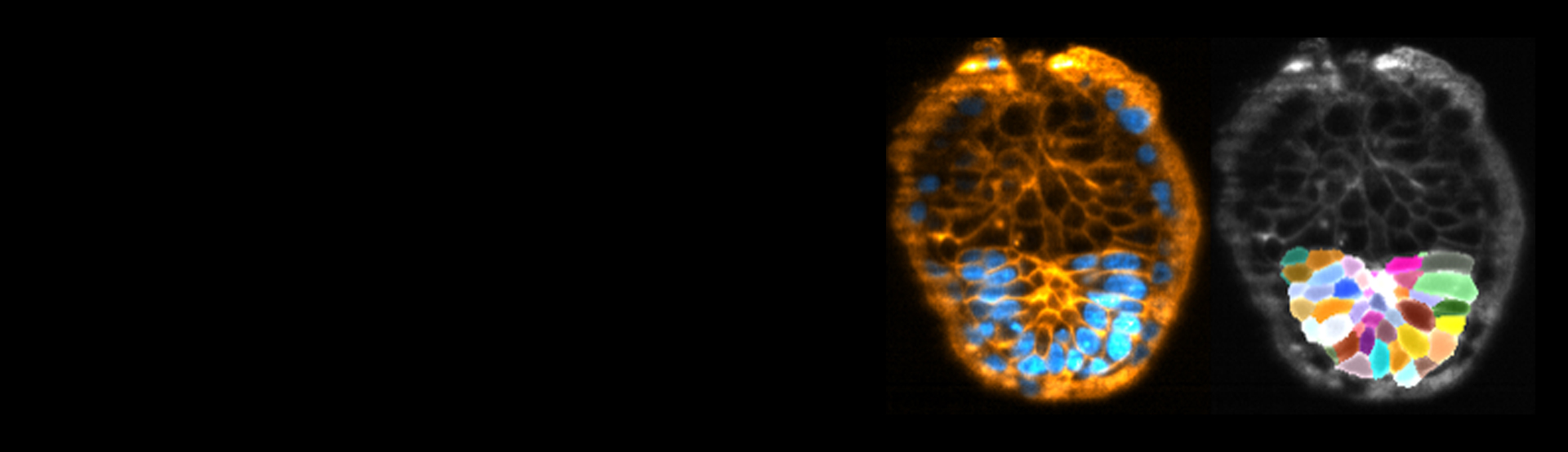
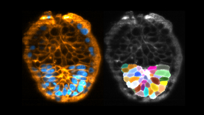
Upon implantation, mammalian embryos undergo major morphogenesis and key developmental processes, such as body axis specification. To gain mechanistic insights into their development, we created a 3D ex vivo culture which not only recapitulates mouse peri-implantation development from E4.5 to E6.0, but also enables us to monitor in toto by using an inverted light-sheet microscope, specifically the InVi SPIM. Our imaging modality accommodates multi-samples for 48 hours of imaging without compromising the development of the embryos, and offers sufficient spatiotemporal resolution for quantitative image analysis, including membrane segmentation. Furthermore, the inverted InVi SPIM has perturbation tools, such as photo-manipulation, that allows us to reveal a key cellular mechanism during peri-implantation development. This ex vivo system will lead to a finer mechanistic understanding of the crucial period of mammalian development.
Find out more about the technology featured in this webinar or our other solutions for light-sheet microscopy:

Featured Products and Technology


Guest Speaker

Takafumi Ichikawa, Ph.D. Assistant Professor, Kyoto University
Takafumi Ichikawa obtained his Ph.D. from Kyoto University in 2017 and undertook postdoctoral training in the Hiiragi group at the European Molecular Biology Laboratory (EMBL) for four years. He was appointed Assistant Professor in 2021 at the Institute for the Advanced Study of Human Biology (ASHBi), Kyoto University. He aims to understand the design principle of tissue patterning and morphogenesis, particularly during mammalian peri-implantation development.
Monika Loeschinger, Senior Product and Applications Specialist, Bruker