
Novel Cancer Drug Target Found Using Electron Paramagnetic Resonance
Researchers at the University of Oxford discovered a novel protein responsible for helping cancer cells survive without oxygen using electron paramagnetic resonance (EPR). This protein may prove to be an important target for future anti-cancer drugs.
Lead author Dr. Iosifina Foskolou and colleagues at the University of Oxford, UK described the findings in their paper, “Ribonucleotide Reductase Requires Subunit Switching in Hypoxia to Maintain DNA Replication,” published in the journal Cell in April of 2017. Using EPR technology, the authors determined the stability and activity of two proteins with important functions in the context of hypoxia to underscore their relative importance in cancer.
Hypoxia, or a lack of oxygen, is a key driver of tumor development and higher levels of hypoxia are associated with worse outcomes in patients.2 Hypoxia speeds up tumor progression by causing replicative stress that leads to increased mutagenesis.3 This helps the cancer cells evade the body’s natural defenses as well as drug therapies.
The authors studied ribonucleotide reductase (RNR), which is a holoenzyme composed of RRM1/RRM2 and RRM1/RRM2B dimers in mammals.4 They found that RRM2B expression is elevated in hypoxic conditions, becoming the dominant subunit in this context. Further, loss of RRM2B reduced cancer cell viability in hypoxic conditions by 77%. The reduction of RRM2B expression increased cancer cell sensitivity to radiological insults, and elevated apoptotic markers were observed specifically in the hypoxic regions of RRM2B depleted tumors.
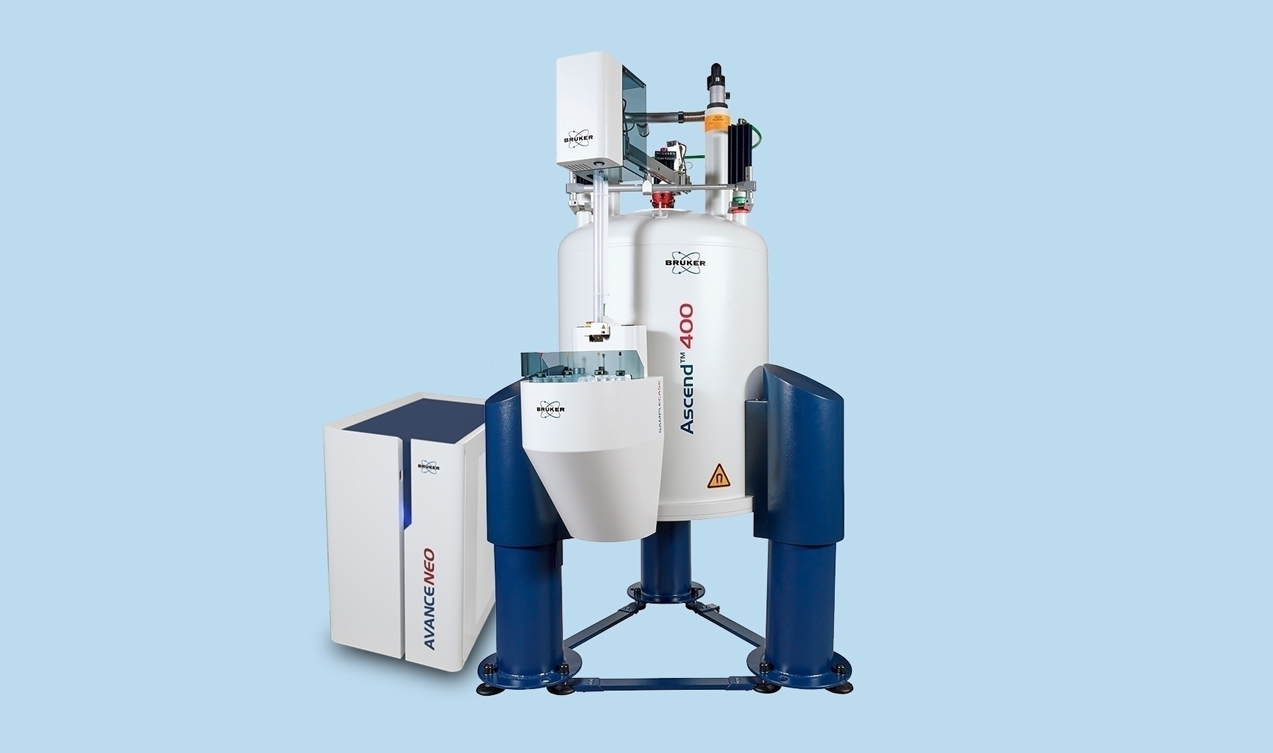
In order to better understand the role of RRM2B in hypoxia, Dr. Foskolou used a Bruker Biospin Micro EMXplus spectrometer to perform EPR studies of RNR subunit stability. The researchers found that RRM2B retains stability and activity under hypoxic stress for up to 2 hours, whereas RRM2 activity is lost after only 15 minutes.
EPR spectrometry, as offered by the Bruker equipment used in this study, is similar to nuclear magnetic resonance (NMR), although it offers increased sensitivity in larger molecules and at shorter time-frames. EPR allows researchers to monitor the dynamic changes in unpaired electrons within a protein, from which the stability can be determined.
By remaining stable and providing the building blocks of DNA, RRM2B allows cancer cells to continue DNA replication in the near absence of oxygen. This, in turn, allows cancer cells to grow and proliferate in an otherwise restrictive environment. Dr. Foskolou and his colleagues hope that by blocking RRM2B, RNR function can be halted in hypoxic conditions.
By determining the amino acid differences between RRM2 and RRM2B that contribute to these divergent activity profiles, the authors have offered insights into moieties that could be targeted by inhibitor molecules.
Whether RRM2B inhibition can improve patient survival in humans with solid tumors remains to be determined. However, the authors found an association between p53-dependent RRM2B expression and hypoxic markers in tumor samples from colorectal adenocarcinoma patients, indicating that these data are relevant to human disease.
“The importance of RRM2B in the response to tumor hypoxia is further illustrated by correlation of its expression with a hypoxic signature in patient samples and its roles in tumor growth and radioresistance,” the authors conclude.
References:
- Foskolou, IP, Jorgensen C, Leszczynska BK, et al. “Ribonucleotide Reductase Requires Subunit Switching in Hypoxia to Maintain DNA Replication.” Mol Cell. 201;66:206–220.
- Begg AC, Stewart FA, Vens C. Strategies to improve radiotherapy with targeted drugs. Nat Rev Cancer. 2011;11:239–253.
- Hammond EM, Kaufmann MR, Giaccia AJ. Oxygen sensing and the DNA-damage response. Curr. Opin Cell Biol. 2007;19:680–684.
- Kolberg M, Strand KR, Graff P, et al. Structure, function, and mechanism of ribonucleotide reductases. Biochim Biophys Acta. 2004;1699:1–34.
- Gunnar Jeschke. Introduction to electron paramagnetic resonance spectroscopy. http://www.chem.ucsb.edu/hangroup/sites/secure.lsit.ucsb.edu.chem.d7_han/files/sitefiles/Links/gunnar-jeschke-lecture_course_updated-1.pdf

