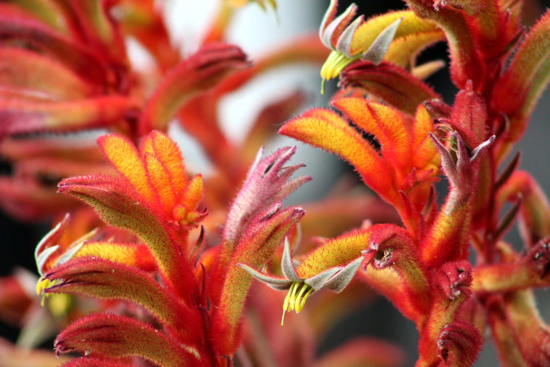

Sugar transport in kangaroo paw
Floral nectar is composed of various components, such as fructose or glucose, which can be visualized using non-specific NMR metabolic profiling. Their distribution is tailored to particular pollinators and thus has a strong influence on the ecosystem. Magnetic resonance microscopy enables tracking the plants metabolism, enabling researches to visualize the formation, transformation, distribution and uptake of the involved compounds to determine the nectar production mechanism
To study the transport of these effects, a magnetic resonance investigation of Anigozanthos, also known as kangaroo paw and catspaw, was carried out. To determine the architecture of tepal nectaries and the connection to the vascular bundle 1H and 13C, images were acquired after labelling the plant in a 500 mM U6-13C glucose solution. The results shown in Figure 2 that the absorbed sugar can be found in the vascular bundles only and is not stored in the tissue
Since the concentration of glucose applied to the stem was very high (500 mM) the enzymatic capacity of the stem was not sufficient to convert all glucose to saccharose. This is the reason why unbound glucose can be observed in the 13C spectrum of the stem, shown in Figure 3.
References: Wenzler, M., Hölscher, D., Oerther, T., & Schneider, B. (2008). Nectar formation and floral nectary anatomy of Anigozanthos flavidus: a combined magnetic resonance imaging and spectroscopy study. Journal of experimental botany, 59(12), 3425-3434.