
Solid-State NMR for Characterization of Bio-Inspired Silica
Solid-state NMR provides a novel approach to the rapid assessment of structural integrity in proteins entrapped in bio-inspired silica
Silica is produced industrially on a huge scale for a range of applications, including catalysis, separations, food and drug technology, biomedical materials and paints. However, this is dwarfed by the volumes of silica deposited in nature. Silica is created by a range of living organisms to provide structural support, protection from predators and optical fibers.
Biosilica, unlike synthetic silica, is produced in an aqueous environment at mild pH and ambient temperatures and has a much higher level of particle organization than synthetic silica. Silica production using natural processes, which are more environmentally friendly and less expensive, has been a key area of development in biotechnology.
Bio-inspired silica is produced through the polycondensation of silicic acid in the presence of a polycationic templating molecule or enzymes. Silica produced in this way can also be used to coat large amounts of protein. Proteins encapsulated in silica are more stable and can be reused. Slight changes in the conditions of synthesis and the type of protein used can give these protein-silica constructs a wide range of morphologies and properties, such as fluorescence or catalytic activity. The possible applications, both medical and industrial, of these biosilica-entrapped proteins are thus endless.
Despite their importance, it has proved difficult to characterize the atomic-level structural of such systems since their size precludes many imaging technologies. Although this has been achieved using solid-state nuclear magnetic resonance (SSNMR), the methodology used relies on 13C detection, which limits the sensitivity, and so a significant sample size is needed for a thorough characterization.
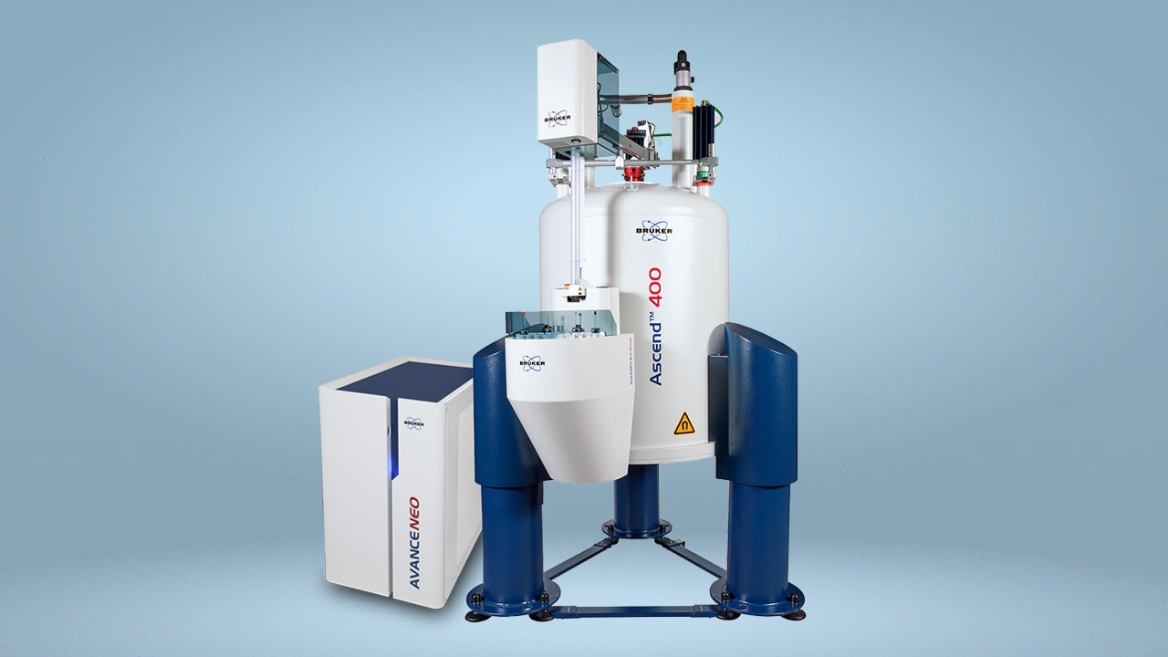
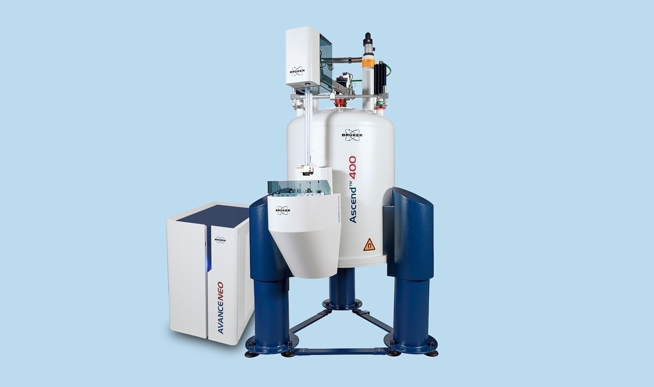
It has recently been shown that the structural integrity in proteins entrapped in bio-inspired silica can be more easily investigated using 1H-detected SSNMR. Although such methodology has already been widely used, this is the first time it has been applied to the assessment of biosilica-entrapped proteins. 1H-15N correlation spectra were obtained for samples of two proteins (green fluorescent protein [GFP] and the catalytic domain of matrix metalloproteinase 12 [catMMP12]), both of which were fused with the biosilica-promoting R5 peptide. All spectra were acquired with a Bruker Avance III spectrometer. Sample packing into Bruker 1.3 mm zirconia rotors was performed with a ultracentrifugal device (courtesy of Bruker Biospin).
Compared with the solution state, most resonances were found to be preserved confirming that the structural integrity of the protein was preserved. The latest approach also offers significant savings in both cost and time, compared with 13C-detection approach. The sample sizes used were significantly lower than those required in previous studies, providing a 9-fold reduction in the cost of sample provision.
Furthermore, the increased sensitivity of the 1H methodology allowed experiment time to be reduced by 4-fold. In addition, the differences in 13C-13C transfers between the solution and solid state mean that more time in required for interpretation of the resulting spectra acquired using 13C methodology compared with spectra obtained with 1H methodology.
Overall, the cost of characterization of biosilica-entrapped proteins using 1H-SSNMR was 80% lower than the equivalent study conducted using the 13C-detection based approach. Use of 1H-SSNMR should facilitate the rapid screening of reaction conditions and/or substrates using a small amount of sample.
References
Ravera E. et al. (1)H-detected solid-state NMR of proteins entrapped in bioinspired silica: a new tool for biomaterials characterization. Sci Rep. 2016;6:27851. © Creative Commons Attribution 4.0 International License: https://creativecommons.org/licenses/by/4.0/


