Application Note: HybridStage™ - Automated, Large Sample-Area Mapping Made Easy
Discover Our Versatile Solutions for Specialized Imaging
The mapping of tissue samples and thicker multi-cellular layers, as well as the nanomechanical characterization of the extracellular matrix and its embedded cells, requires a (semi)automated AFM approach for a millimeter range in x,y with nm–resolution. Large variations in the topography of tissue samples, combined with fluidity and stickiness of membrane and extracellular proteins, require greater flexibility in the z-axis of the AFM.
Our HybridStage—a modular, piezo-based sample scanner stage combined with motorized XY sample movement—provides a highly versatile solution to these challenges.
The HybridStage is a tool for advanced, multi-parametric AFM characterization of samples in the range of mm to nm and pN resolution, which features three scanner units that can move the tip/sample. A wide range of samples can be studied, including biomaterials, cells and macroaggregates, embryos and tissues, model organisms in developmental biology (zebrafish, C. elegans, etc), and implants. Particularly the increased z-piezo range, equipped with wide-ranged motorized sample motion, is perfect for the nanomechanical characterization of tissues required in cancer, nanomedicine, or developmental biology.
Readers can expect to learn about:
- The capabilities of the HybridStage;
- The advantages of large-range tiling of optical images; and
- How the HybridStage, together with NanoWizard software, facilitates multi-scan force mapping over large areas on very rough, very soft, and sticky samples.
KEYWORDS: Cell-Cell Interactions; Extracellular Matrix Proteins; Optical Tiling; Single Cell Force Spectroscopy; Tissue Mapping

Introduction
Crucial parameters that affect cell adhesion, morphogenesis, cell differentiation and cancer invasion include the molecular interactions between cells and their extracellular matrix environment, their 3D topography and the corresponding mechanical properties. 1-3. AFM (atomic force microscopy) is an advanced multi-parametric imaging technique which delivers 3D profiles of the surfaces of molecules and cells in the nm-range. It also enables the characterization of nanomechanical properties (adhesion, elasticity etc.) and the visualization of structural changes taking place at the molecular level.


The mapping of tissue samples and thicker multi-cellular layers as well as the nanomechanical characterization of the extracellular matrix and its embedded cells requires a (semi)automated AFM approach for a millimeter range in x,y with nm-resolution. Large variations in the topography of tissue samples, combined with fluidity and stickiness of membrane and extracellular proteins, require a larger flexibility in the z-axis of the AFM 3.
Technical solution
The JPK BioAFM Center has developed a highly versatile solution: HybridStage. It is equipped with three scanner units that can move either the tip or the sample. A standard NanoWizard AFM head has an xyz tip scanner with a scan range of 100 x 100 x 15 µm3. The HybridStage houses an xyz sample scanner with an optional scan range of either 100 x 100 x 100 µm3, 200 x 200 x 200 µm3 or 300 x 300 x 300 µm3, which can be individually selected depending on the individual application. It also features a motorized unit for large sample movements (20 x 20 mm2). The wide choice of scanners, together with optical tiling and multi-region AFM probing (figures 6, 7), enables multiparametric characterization of soft samples over a large area, and provides additional optical data sets on inverted and upright microscopes respectively (figures 1, 2).
Single Cell Force Spectroscopy and membrane tethers
The most frequent activities in the field of Single Cell Force Spectroscopy (SCFS) are the study of the adhesion of individual cells on (bio )materials, extracellular matrix proteins and the characterization of cell-cell interactions 4.
Due to the effect of membrane tethering, the pulling length required to separate cells from a substrate frequently exceeds several 10th5 of microns 5. The flexible, modular design of the HybridStage, with 3D sample scanners that can scan in the range of 100 x 100 x 100 µm3, 200 x 200 x 200 µm3 or 300 x 300 x 300 µm3 can meet this demand. Figure 3 illustrates a detachment curve of a fibronectin functionalized cantilever from a Vero cell. After reaching the cell surface at a piezo height of approx. 60 µm, separation is completed at a final piezo height of 105 µm. The membrane tube formations, with force plateaus of 1 - 15 µm in length, are the main reason for a total separation distance of about 45 µm in this example.

Optical focus control with JPK Slim FocusTM
The consequence of a long-ranged z-movement for the tip and sample scanner is that either the cantilever or the sample move out of the optical focus. Therefore, we have implemented the JPK SlimFocus (Figure 4) to synchronize the objective lens motion with the current z-scanner (figure 5).
The JPK SlimFocus control software allows manual adjustment of the z-position using a slider, and the setting of two marker positions, e. g. for the tip/sample. During AFM experiments, the optical focus motion can be synchronized with the z-scanner currently being used up to 150 µm.


Optical Tiling and Multi-Region Imaging
If the x,y piezo scan range and the optical observation window (typically 100 µm) are much smaller than the sample size (in the range of mm) to be investigated with the AFM, a conflict arises. In order to enlarge the microscopic optical overview, the motorized sample scanner executes a repetitive pattern, acquiring the corresponding optical images of the sample which are then tiled and displayed in the SPM software (Figure 6). When tiling starts, the file chooser opens an automatically named sub-folder. The calibration file and the image file are saved in the sub-folder and are easily accessible for data processing.


For multi-region AFM imaging, the user will be asked whether they wish to examine more than one measurement position. They can then select points of interest before a measurement starts, which will then be displayed in the data viewer with tiled optical images. When the measurement begins, the positions are processed sequentially. Different types of measurement definitions are available: single points, lines and rectangular regions. The multi-scan option is available for most measurement modes like force mapping and spectroscopy, AFM imaging, QI™ and multiple DirectOverlay TM snapshots. An example of multiscan QI imaging of mammalian Vero cells on an inverted optical microscope can be seen in Figure 7. First, DirectOverlay was performed to correlate the cantilever tip position with the corresponding optical image. Second, optical tiling with 4 x 4 images on 500 x 500 µm2 was carried out (see figure 6) to increase the region of interest. Third, a multi-scan range was selected. For this specific example, 4 x 4 QI scans, with an individual map size of 50 µm, were performed (frames are labeled in magenta). In Figure 7, 13 of the 16 maps have already been executed. The green frame on position 14 indicates the current map position. Using the "advanced force oscilloscope", multiple data viewers can be activated, e.g. height (of the sample) at force setpoint, adhesion (between cantilever and sample), slope (as an indication of sample stiffness) and reference force height(s) at a defined value of the force setpoint. In the specific example shown in Figure 7, two different data viewer channels are displayed (height at 100% of the setpoint and reference height at 80% of the setpoint). To obtain a better contrast of cellular structures, the reference height is indicated as a pixel difference image.
Multi-Scan Tissue Mapping
In order to obtain a better understanding of the development and progression of cancer, analyzing changes in the mechanical properties of cells and their surrounding extracellular matrix material appears to be crucial 3. Using the example of human cervix tissue, the benefits of the HybridStage and the mode of operation, in combination with an upright macroscope, are presented here.


Thanks to its long working distance, the upright macroscope allows the collection of optical images with or without the AFM head. Furthermore, replacement of the AFM head does not require a readjustment of the sample. The excellent replacement accuracy of the head with a mounted cantilever is in the range of< 2µm in x,y,z. Once the region of interest is found in the optical image, it can be used to position the AFM probe with respect to the area of interest (Figure 8). A HybridStage with a 3D piezo sample scanner of 300 x 300 x 300 µm3 in conjunction with the motorized stage was used to perform a multi-scan map of 1000 x 1000 µm2 with 5 x 5 single maps of 200 x 200 µm2 and a pixel distance of 10 µm each (Figure 9). All individual maps were analyzed using the batch processing option for indentation experiments in the JPK data processing (DP) software. Alternatively, the data can be exported, analyzed and composed using a third-party software e. g. Matlab. Figure 10 summarizes the frequency distribution of the apparent Young's modulus of the multi-scan shown in Figure 9.
When performing a multi-scan force map, over a larger area on a very rough sample with varying heights, it may e. g. i) damage the cantilever, or ii) the AFM measurements may not yield usable data. Furthermore, in very soft samples with sticky surfaces, the cantilever only separates from the sample surface after several 10th of micrometers, resulting in noticeable deformation of the sample or removal of the membrane tube.


Here too, the z-range must be increased in order to successfully complete the measurement. To solve this technical difficulty, the NanoWizard software can set the height of both the piezo-retract and the motor-retract. In this way, an individual map and entire scan can be optimally performed. Figure 11 shows that the tissue sample examined is very rough and has a very varied height. Even in the individual map of 200 x 200 µm2, the height difference is approx. 80 µm. Despite this, the multi-scan could still stably perform the measurement without having to stop.
HybridStage Compatibilities
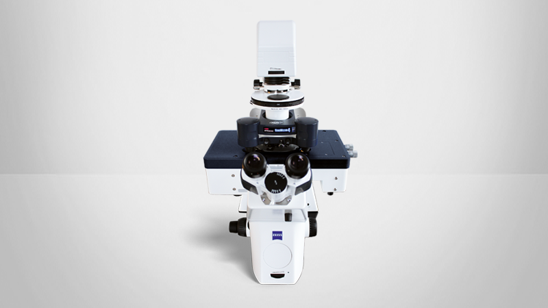
Optical microscope: Upright
- Zeiss Axio Zoom.V16
- Leica MF205
- Olympus MVX 10 with epi-fluorescence and brightfield
Optical microscope: Inverted
- Zeiss AxioObserver
- Nikon TE, Ti and Ti2 lines
- Leica DMI 6000/DMi8
- Olympus IX71 with epi-fluorescence and brightfield
Conclusion
The HybridStage is a versatile tool for advanced, multiparametric AFM characterization of samples in the range of mm to nm and pN resolution, which features three scanner units that can move the tip/sample. A wide range of samples can be studied, including biomaterials, cells and microaggregates, embryos and tissues, model organisms in developmental biology (zebra-fish, C. elegans, etc) and implants. Particularly the increased z-piezo range, equipped with wide-ranged motorized sample motion, is perfect for the nanomechanical characterization of tissues required in cancer, nanomedicine or developmental biology.
The HybridStage frees experiments from the constraints of the AFM piezo range. Large-range tiling of optical images provides a clear visual overview, allowing a fast setup of optically guided experiments and direct selection of the optical features for investigation. Navigate around the sample, collect a list of interesting features for multi-scan, or even map force responses over greatly extended scan ranges. The HybridStage is a modular, piezo-based sample scanner stage combined with motorized XY sample movement, giving direct access to anything you can see.
Sample courtesy
The data for tissue characterization shown in Figures 2 and 8 - 11 are kindly provided by Prof. J. Käs and Dr. Th. Fuhs (University Leipzig). The Vero cell samples were provided by Prof. A. Hermann and his group (Humboldt university Berlin).
References
- R. Pearce, G. J. Vansco, Macromolecules, 30: 5853 (1997)
- T.J. McMaster, J.K. Hobbs, P.J. Barham, M.J. Miles, Probe Microsc., 1: 43 (1997)
- J.K. Hobbs, T.J. McMaster, M.J. Miles, P.J. Barham, Polymer, 39: 2437 (1998)
- K.K. Jain, Drug Delivery Systems, Springer, 2008
- V.H. Mareau, R.E. Prud'homme., Macromolecules, 38: 398 (2007)
- V. Speranza, A. Sorrentino, F. De Santis, R. Pantani, Hindawi Publishing Corporation- The Scientific World Journal, (2014), Article ID 720157, http://dx.doi.org/10.1155/2014/720157
- sample courtesy Nie Mullin, University of Sheffield, UK
- https://www.youtube.com/user/JPKlnstruments
- E. Zhuravlev, PhD thesis, Universtiy of Rostock, Germany, http://www.polymerphysik.unirostock.de/publication/theses/pdf/Zhuravlev .pdf