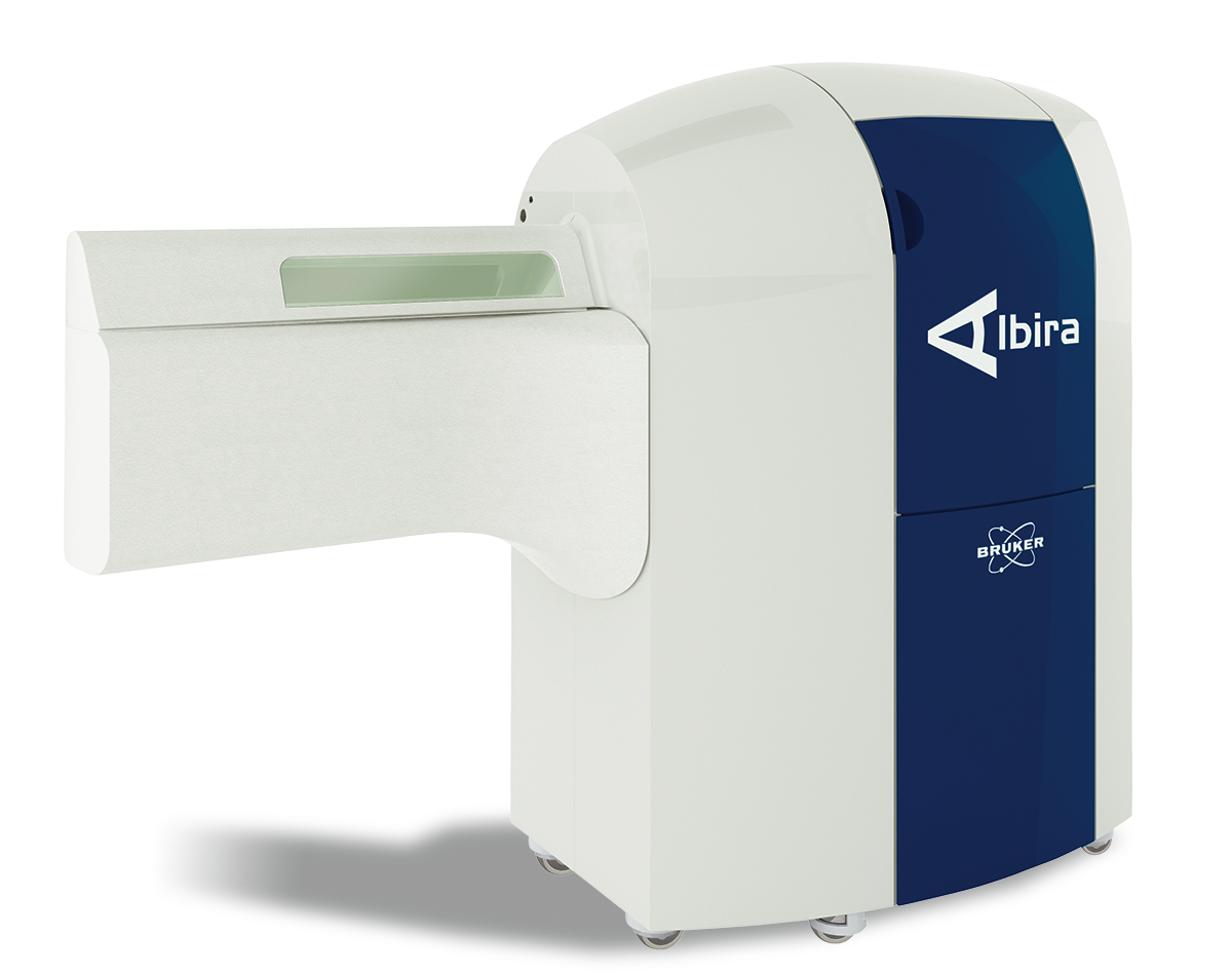
Albira SI
Truly multimodal
highly flexible


Like you, thousands of our customers around the world rely on advanced imaging techniques to study dynamic biological process within living animals. To better understand the progression and treatment of diseases, you can use Albira's PET, SPECT and CT imaging for quantitative 3D tomographic imaging of radiotracers, bone, and soft tissue.
Features
Animal Care Monitoring And Control
- Anesthesia: Fully integrated. Compatible with most common commercial gas systems
- Animal Handling System: Reliable, easy to use rat and mouse beds; Fully compatible with BRUKER MR
- Physiological Signals: ECG, Respiration, Temperature, And Blood Pressure
- Temperature Control System: Electrically heated mats for rat And mouse
- Video Monitoring: Realtime camera
- Gating Acquisition: Cardiac and Respiratory For PET and SPECT; Dual Gating for PET
Software and Workstation
- Fully Integrated Albira Suite: ACQUIRER: Image Acquisition, RECONSTRUCTOR: Image Reconstruction, MANAGER: Study and Protocol Management, SUPERVISOR: Quality Control
- Image Analysis Software: PMOD (PBAS, PFUS, PKIN MODULES INCLUDED)
- Workstation: Dedicated Server; All Functionalities on a single system (Acquisition, Storage, Reconstruction and Image Processing)
- Reconstruction: GPU Based, CPU Based SPECT
68Ga Tracer Comparison in Tumor Mice
Micro PET/CT allows comparison of [68Ga]FSC (succ-RGD)3 with [68Ga]NODAGA-RGD in a human glioblastoma model (two mice)
3D volume rendered projections of fused microPET/CT static images for [68Ga]FSC(succ-RGD)3 and [68Ga]NODAGA-RGD at 90 min p.i.
Courtesy: Institute of Molecular and Translational Medicine, Palacky University, Olomouc, Czech Republic.
Reference: Zhai C, Franssen GM, Petrik M, et al. Molecular Imaging and Biology. 2016;18(5):758-767. doi:10.1007/s11307-016-0931-3.
Dynamic 18F-FDG PET Imaging
In vivo dynamic PET imaging and kinetic modeling supports full quantification of myocardium glucose metabolism.
Courtesy: B Kundu, M Chordia, J Li and S Berr, University of Virginia, US
68Ga Biodistribution Studies
Static microPET/CT images of [68Ga]-AAZTA-MG in CCK+/−, tumour xenograft BALB/c nude mice. 30 (A, D), 45 (B, E) and 60 (C, F) min p.i.; coronal slices (A, B, C) and 3D volume rendered images (D, E, F)
Courtesy: Institute of Molecular and Translational Medicine, Palacky University, Olomouc, Czech Republic.
Reference: Pfister J, Summer D, Rangger C, et al. EJNMMI Research. 2015;5:74. doi:10.1186/s13550-015-0154-7.
Tumor Specific Targeting Using 68Ga Bombesin
High sensitivity PET imaging and automatic CT co-registration allows for an accurate evaluation of drug efficacy mechanisms.
Courtesy: D. Viertl, F. Cicone & J. Prior, D. Viertl. CHUV Lausanne
Effect of Neurodegenerative Therapy
TgF344-AD rat model imaged with AMYViD-PET/CT and co-registered with brain atlas for regional measurements.
PET imaging reveals marked reduction in Aβ levels at pituitary, auditory cortex visual cortex and ventral tegmental in treated rats.
Courtesy: S Berr, University of Virginia Radiology and Biomedical Engineering and B Costa, Virginia College of Osteopathic Medicine.
PET and MR in Mouse MetastaticTumor Model
(Left) Coronal whole-body PET/MR. (Right) Focused Axial PET/MR revealing location of lung and abdominal tumors.
Courtesy of Acknowledgements. Thanks to the Katherine Tucker, Mikayla Rodger, Dr. Lixin Ma, Dr. Carolyn Anderson, and Dr. Barry Edwards, MITC – Molecular & Imaging Theranostics Center, University of Missouri.
Total body PET/MRI images of mice
Imaged at pre-injection and 1, 5, 30, 45 mins post-injection of 0.1 mmol/kg [52Mn]Mn-2CHyd or [52Mn]Mn-1CHyd.
Courtesy Yingying Ning , Iris Yuwen Zhou, Nicholas J Rotile et al. MGH – USA
Reference: J Am Chem Soc. 2022 Sep 14;144(36):16553-16558.
64Cu Tracer Validations/Characterizations in a Mouse Glioblastoma Model
AD Model in Marmosets: Awake Imaging with 11C-PiB and 18F-FDG PET
PET/CT Si78
Specifications
- Imaging Modalities: PET, SPECT, CT
- Coincidence based PET featuring Full Field of View Accuracy which means constant resolution across the entire FOV for consistent quantification
- Adaptative SPECT FOV from to 25 to 120 mm (in one single bed position) and 4 different collimator types
- Single compact footprint 90 × 220 cm
- Fully Shielded against X-rays
Software
Webinars
Testimonials
Learn More
Read more on Albira Si:
Application Notes
- Advantages of Integrated Hybrid Imaging
- Advantages of Coincidence PET
- Multi-modal PET drives interdisciplinary preclinical imaging
- Setting up a Preclinical Imaging Lab
- Considerations on Exposure to Radiation in a Preclinical Lab
- Blood Sampling in Preclinical PET
- PET Imaging Studies of Candidate Therapeutics
- Imaging Cancer: Multi-modal PET Technology Provides Novel Insights in Preclinical Oncology
- The Use of Positron Emission Tomography (PET) in Preclinical Pharmaceutical Research
LabScape
Service & Life Cycle Support for Magnetic Resonance and Preclinical Imaging
Bruker’s commitment to provide customers with unparalleled help throughout the buying cycle, from initial inquiry to evaluation, installation, and the lifetime of the instrument is now characterized by the LabScape service concept.
LabScape Maintenance Agreements, On-Site On-Demand and Enhance Your Lab are designed to offer a new approach to maintenance and service for the modern laboratory
