
Sequence-Specific Assignments for Intrinsically Disordered Proteins
At present, we do not fully understand the function, structure, and dynamics of intrinsically disordered proteins (IDP) in biological systems.
Due to their characteristic lack of structure and high mobility, analyzing IDPs can be difficult, but nuclear magnetic resonance (NMR) can provide sequence-specific information about these fascinating macromolecules.1-3
Proline signals are usually invisible in 1H-15N NMR experiments because they lack a hydrogen atom on their alpha-amino group. However, they can play an essential role in maintaining a flexible structure and preventing the formation of secondary motifs in IDPs.4
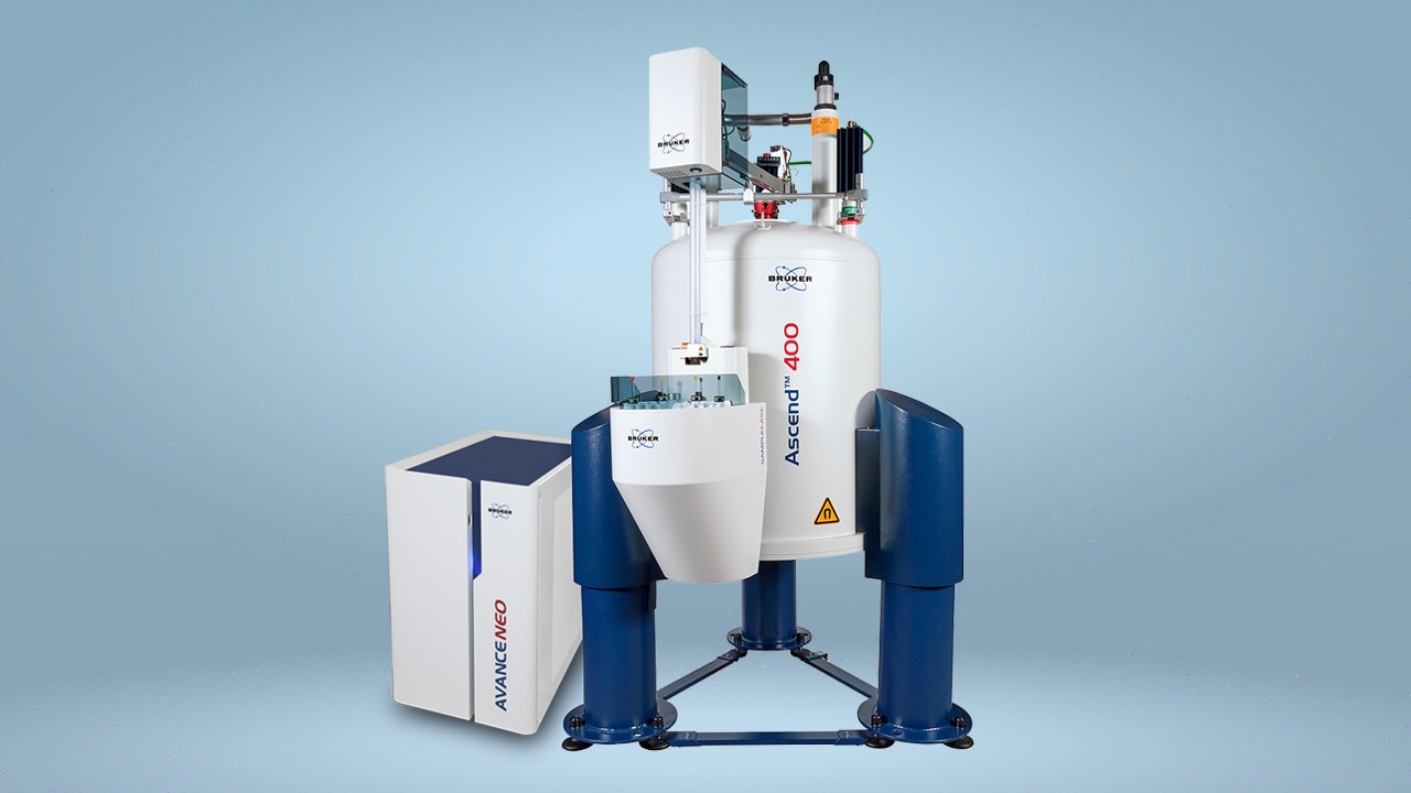
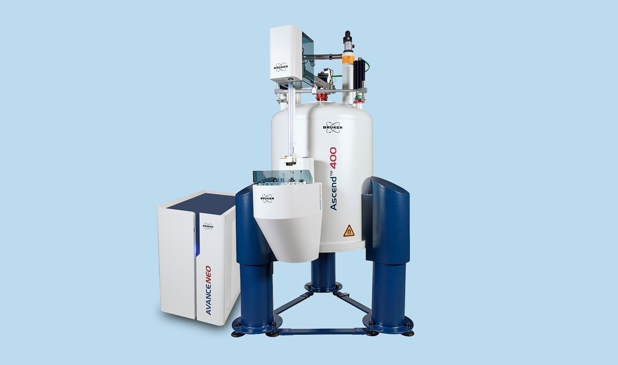
A recent publication in ChemBioChem details how researchers from Italy used 13C direct‐detection NMR spectroscopy to detect proline residues in IDPs. Their NMR spectra were acquired on a Bruker AVANCE NEO spectrometer equipped with a cryogenically cooled probe head optimized for 13C-direct detection.4
Using their technique, they were able to produce a proline fingerprint spectrum for an IDP called CBP-ID4, successfully detecting all 45 proline residues in the protein, with almost no spectral overlap. 4
The NMR spectra of IDPs
3D triple-resonance experiments using 13C, 15N, and 1H often allow the complete assignment of NMR signals from globular proteins. However, the flexibility of IDPs means that 3D triple resonance does not provide enough resolution for complete assignment, and high resolution in multi-dimensions is required.3,5,6
A publication in the Journal of Biomolecular NMR by the same team of Italian researchers outlines how they used 13C-direct detected APSY NMR experiments to achieve the sequence-specific assignment of alpha-synuclein.6
They used the same Bruker AVANCE NEO spectrometer and probe set up as in their previous study, and they were able to assign the chemical shifts of C’ and N atoms in peptide bonds along the backbone of alpha-synuclein, obtaining information on the amino acid sequence along the protein backbone.6
Detecting post-translational modifications
IDPs, like all proteins, can be subjected to a range of post-translational changes. These modifications can be challenging to detect and study using NMR. A recent paper by scientists from Masaryk University in the Czech Republic describes how they used 1D 31P NMR to detect the presence of phosphorylation modifications in IDPs, and 2D 1H-13C, 1H-15N HSQC experiments to identify the phosphorylated residues. Their NMR spectra were obtained with a Bruker AVANCE III HD Spectrometer (Now AVANCE NEO)7.
Their 31P spectra successfully confirmed two phosphorylation sites in their model peptide, while HSQC experiments identified tyrosine as the phosphorylated residue in their model protein. 7
Conclusions
NMR is a powerful technique for studying the structure and dynamics of IDPs. New techniques can overcome the challenges of incomplete assignment, low chemical shift dispersion, and detecting post-translational modifications in IDPs. Characterizing biologically relevant IDPs could aid the development of new therapies in a range of diseases.
References
- ‘Structure and Function of Intrinsically Disordered Proteins’ —Tompa P, Fersht A, CRC, 2009.
- ‘Application of NMR to studies of intrinsically disordered proteins’ — Gibbs EB, Cook EC, Showalter SA, Archives of Biochemistry and Biophysics, 2017.
- ‘NMR contributions to structural dynamics studies of intrinsically disordered proteins’ — Konrat R, Journal of Magnetic Resonance, 2014.
- ‘Prolines’ fingerprint in intrinsically disordered proteins’ — Murrali MG, Piai A, Bermel W, Felli IC, Pierattelli R, ChemBioChem, 2018.
- ‘Intrinsically Disordered Proteins Studied by NMR Spectroscopy’ —Felli IC, Pierattelli R, Springer, 2015.
- ‘13C APSY-NMR for sequential assignment of intrinsically disordered proteins’ — Murrali MG, Schiavina M, Sainati V, Bermel W, Pierattelli R, Felli IC, Journal of Biomolecular NMR, 2018.
- ‘Efficient and robust preparation of tyrosine phosphorylated intrinsically disordered proteins’ —Brázda P, Sedo O, Kubícek K, Štefl R, BioTechniques, 2019.


