
Advances in Light-Sheet Microscopy: A Universal Tool for 3D Bioimaging
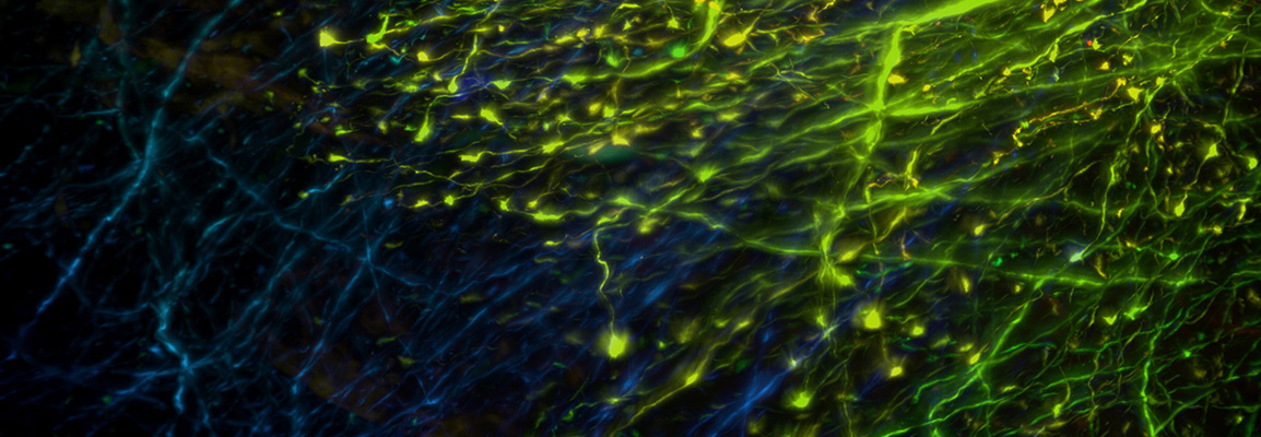
Light-sheet microscopy is the coming-of-age fluorescence imaging technique in fields as diverse as cell and developmental biology, neuroscience, oncology, plant research, high-content screening or biophysics. It enables long-term and large-scale 3D imaging at much higher imaging speed but without the inevitable phototoxicity or photobleaching that occurs when using fluorescence imaging techniques like laser scanning or spinning disk confocal microscopy.
In this webinar, we will discuss the benefits of light-sheet fluorescence microscopy as a gentle bio-imaging technique and present an overview of our latest product developments. We will show you how you can use the Luxendo light-sheet microscopes MuVi SPIM, InVi SPIM, TruLive3D Imager and QuVi SPIM for imaging of developing embryos or 2D and 3D culture systems like cell lines and organoids under full environmental control. In addition, we will also cover the application of MuVi SPIM, QuVi SPIM and LCS SPIM for 3D microscopy of cleared samples. Whether you need high resolution, which so far has only been achieved using confocal microscopy, or whether you aim at imaging large objects like entire mouse or rat brains or organs – we have an offering. Finally, we will describe our innovative and versatile sample mounting techniques, including our disposable TruLive3D Dishes.
What to Expect
In this webinar, you can expect to learn the advantages of light-sheet fluorescence microscopy and how this technique can be adapted to a multitude of different live, fixed or cleared samples and image these without the phototoxicity that plagues traditional imaging techniques. Expect to see an overview of Bruker’s range of Luxendo light-sheet microscopes.
Key Topics
- Learn about the advantages of light-sheet fluorescence microscopy
- How to use it for a multitude of different live, fixed or cleared samples
- Get an overview of Bruker’s Luxendo light-sheet microscopes
Speaker
Dr. Malte Wachsmuth
Head of Application, Support and Service at Luxendo GmbH, Bruker Nano Group
Dr. Malte Wachsmuth is a Co-founder and Head of Applications, Support & Service at Luxendo GmbH. His extensive research and business knowledge in the field of Light-Sheet Fluorescence Microscopy is based on a solid scientific career developed over the last years. After graduating from the University of Hamburg, he completed a Ph.D. in Biophysics at the German Cancer Research Center (DKFZ), in Heidelberg. Additionally, he held leading positons in renowned research centers such as the Cell Biophysics Group at Institut Pasteur in Korea and the Cell Biology & Biophysics Unit at European Molecular Biology Laboratory (EMBL), in Heidelberg, Germany.


