Languages
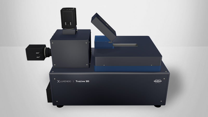
Imaging Organoids with Light-Sheet Fluorescence Microscopy
Explore the Advantages of Light-Sheet Microscopy for Organoid Research

When imaging organoids, collecting high-quality images requires striking a balance between acquisition time, sample size and field-of-view requirements, and the duration of the experiment. Light-sheet fluorescence microscopy (LSFM) rises to this challenge, allowing researchers to increase acquisition speed, decrease phototoxicity, and obtain high-resolution, low-noise images of organoid samples. Moreover, by enabling gentle three-dimensional imaging of organoids—and even their cellular structures—with limited phototoxicity and fast acquisition, LSFM provides several advantages over other methods of imaging organoids and opens new frontiers in organoid research.
Readers can expect to learn more about:
- The unique capabilities of light-sheet fluorescence microscopy;
- Its advantages for imaging organoids; and
- The innovative research it now supports.
This brief includes a case study showcasing the use of light-sheet microscopy for organoid imaging in cancer biology research.
KEYWORDS: Cancer Biology Research; Case Study; Light-Sheet Microscopy; Organoids; Phototoxicity